5NYW | pdb_00005nyw

Anbu (ancestral beta-subunit) from Yersinia bercovieri


Experimental Data Snapshot

  • Method: X-RAY DIFFRACTION
  • Resolution: 2.50 Å
  • R-Value Free: 
    0.230 (Depositor), 0.231 (DCC) 
  • R-Value Work: 
    0.183 (Depositor), 0.189 (DCC) 
  • R-Value Observed: 
    0.183 (Depositor) 

Starting Model: experimental
View more details

wwPDB Validation 3D Report Full Report

Validation slider image for 5NYW

This is version 2.0 of the entry. See complete history

Literature

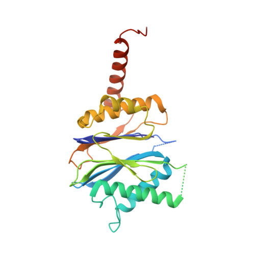
The Y. bercovieri Anbu crystal structure sheds light on the evolution of highly (pseudo)symmetric multimers.

Piasecka, A.Czapinska, H.Vielberg, M.T.Szczepanowski, R.H.Kiefersauer, R.Reed, S.Groll, M.Bochtler, M.

(2018) J Mol Biology 430: 611-627

  • DOI: https://doi.org/10.1016/j.jmb.2017.11.016
  • Primary Citation Related Structures: 
    5NYW

  • PubMed Abstract: 

    Ancestral β-subunit (Anbu) is homologous to HslV and 20S proteasomes. Based on its phylogenetic distribution and sequence clustering, Anbu has been proposed as the "ancestral" form of proteasomes. Here, we report biochemical data, small-angle X-ray scattering results, negative-stain electron microscopy micrographs and a crystal structure of the Anbu particle from Yersinia bercovieri (YbAnbu). All data are consistent with YbAnbu forming defined 12-14 subunit multimers that differ in shape from both HslV and 20S proteasomes. The crystal structure reveals that YbAnbu subunits form tight dimers, held together in part by the Anbu specific C-terminal helices. These dimers ("protomers") further assemble into a low-rise left-handed staircase. The lock-washer shape of YbAnbu is consistent with the presence of defined multimers, X-ray diffraction data in solution and negative-stain electron microscopy images. The presented structure suggests a possible evolutionary pathway from helical filaments to highly symmetric or pseudosymmetric multimer structures. YbAnbu subunits have the Ntn-hydrolase fold, a putative S 1 pocket and conserved candidate catalytic residues Thr1, Asp17 and Lys32(33). Nevertheless, we did not detect any YbAnbu peptidase or amidase activity. However, we could document orthophosphate production from ATP catalyzed by the ATP-grasp protein encoded in the Y. bercovieri Anbu operon.


  • Organizational Affiliation
    • Polish Academy of Sciences, Institute of Biochemistry and Biophysics, Warsaw, Poland; School of Medicine, Cardiff University, Cardiff, United Kingdom.

Macromolecule Content 

  • Total Structure Weight: 790.13 kDa 
  • Atom Count: 52,945 
  • Modeled Residue Count: 6,523 
  • Deposited Residue Count: 7,028 
  • Unique protein chains: 1

Macromolecules

Find similar proteins by:|  3D Structure
Entity ID: 1
MoleculeChains  Sequence LengthOrganismDetailsImage
Peptidase
A,
AA [auth 1],
B,
BA [auth 2],
C,
D,
E,
F,
G,
H,
I,
J,
K,
L,
M,
N,
O,
P,
Q,
R,
S,
T,
U,
V,
W,
X,
Y,
Z
251Yersinia bercovieriMutation(s): 0 
Gene Names: CS533_03425
UniProt
Find proteins for A0A2G4U6U6 (Yersinia bercovieri)
Explore A0A2G4U6U6 
Go to UniProtKB:  A0A2G4U6U6
Entity Groups
Sequence Clusters30% Identity50% Identity70% Identity90% Identity95% Identity100% Identity
UniProt GroupA0A2G4U6U6
Sequence Annotations
Expand
Reference Sequence

Small Molecules

Ligands 6 Unique
IDChains Name / Formula / InChI Key2D Diagram3D Interactions
PEG

Query on PEG


Download:Ideal Coordinates CCD File
IB [auth T],
XA [auth N]
DI(HYDROXYETHYL)ETHER
C4 H10 O3
MTHSVFCYNBDYFN-UHFFFAOYSA-N
SO4

Query on SO4


Download:Ideal Coordinates CCD File
AB [auth P]
CA [auth A]
DA [auth B]
EA [auth B]
KA [auth E]
AB [auth P],
CA [auth A],
DA [auth B],
EA [auth B],
KA [auth E],
LA [auth E],
LB [auth V],
MB [auth W],
OB [auth X],
PB [auth X],
QB [auth X],
RB [auth Z],
TB [auth 1],
UA [auth L],
VB [auth 2],
WB [auth 2],
ZA [auth P]
SULFATE ION
O4 S
QAOWNCQODCNURD-UHFFFAOYSA-L
EDO

Query on EDO


Download:Ideal Coordinates CCD File
FA [auth C]
HA [auth D]
MA [auth F]
QA [auth K]
TA [auth L]
FA [auth C],
HA [auth D],
MA [auth F],
QA [auth K],
TA [auth L],
YA [auth O]
1,2-ETHANEDIOL
C2 H6 O2
LYCAIKOWRPUZTN-UHFFFAOYSA-N
AZI

Query on AZI


Download:Ideal Coordinates CCD File
PA [auth J]AZIDE ION
N3
IVRMZWNICZWHMI-UHFFFAOYSA-N
CL

Query on CL


Download:Ideal Coordinates CCD File
BB [auth P]
CB [auth Q]
FB [auth R]
GA [auth C]
GB [auth S]
BB [auth P],
CB [auth Q],
FB [auth R],
GA [auth C],
GB [auth S],
HB [auth T],
IA [auth D],
JA [auth D],
JB [auth U],
NA [auth G],
NB [auth W],
RA [auth K],
SA [auth K],
SB [auth Z],
UB [auth 1],
VA [auth L],
WA [auth M]
CHLORIDE ION
Cl
VEXZGXHMUGYJMC-UHFFFAOYSA-M
UNX

Query on UNX


Download:Ideal Coordinates CCD File
DB [auth Q],
EB [auth Q],
KB [auth U],
OA [auth I]
UNKNOWN ATOM OR ION
X

Experimental Data & Validation

Experimental Data

  • Method: X-RAY DIFFRACTION
  • Resolution: 2.50 Å
  • R-Value Free:  0.230 (Depositor), 0.231 (DCC) 
  • R-Value Work:  0.183 (Depositor), 0.189 (DCC) 
  • R-Value Observed: 0.183 (Depositor) 
Space Group: P 1 21 1
Unit Cell:
Length ( Å )Angle ( ˚ )
a = 95.535α = 90
b = 285.371β = 91.78
c = 179.213γ = 90
Software Package:
Software NamePurpose
PHENIXrefinement
XDSdata reduction
XSCALEdata scaling
PHASERphasing
Cootmodel building
ARP/wARPmodel building

Structure Validation

View Full Validation Report


Entry History 

& Funding Information

Deposition Data


Funding OrganizationLocationGrant Number
National Science CentrePolandNCN UMO-2013/08/S/NZ1/00681
Cardiff UniversityUnited Kingdom--

Revision History  (Full details and data files)

  • Version 1.0: 2017-05-24
    Type: Initial release
  • Version 1.1: 2017-06-28
    Changes: Refinement description
  • Version 1.2: 2018-01-03
    Changes: Database references
  • Version 1.3: 2018-03-07
    Changes: Database references
  • Version 1.4: 2024-01-31
    Changes: Data collection, Database references, Refinement description
  • Version 2.0: 2025-02-05
    Changes: Advisory, Atomic model, Data collection, Database references, Derived calculations, Non-polymer description, Refinement description, Source and taxonomy, Structure summary