5NX9 | pdb_00005nx9

Crystal structure of Neanderthal Adenylosuccinate Lyase (ADSL) in complex with its products AMP and fumarate


Experimental Data Snapshot

  • Method: X-RAY DIFFRACTION
  • Resolution: 2.30 Å
  • R-Value Free: 
    0.227 (Depositor), 0.193 (DCC) 
  • R-Value Work: 
    0.193 (Depositor) 
  • R-Value Observed: 
    0.195 (Depositor) 

Starting Model: experimental
View more details

wwPDB Validation 3D Report Full Report

Validation slider image for 5NX9

Ligand Structure Quality Assessment 


This is version 2.1 of the entry. See complete history

Literature

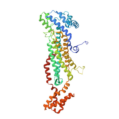
Molecular comparison of Neanderthal and Modern Human adenylosuccinate lyase.

Van Laer, B.Kapp, U.Soler-Lopez, M.Moczulska, K.Paabo, S.Leonard, G.Mueller-Dieckmann, C.

(2018) Sci Rep 8: 18008-18008

  • DOI: https://doi.org/10.1038/s41598-018-36195-5
  • Primary Citation Related Structures: 
    5NX8, 5NX9, 5NXA

  • PubMed Abstract: 

    The availability of genomic data from extinct homini such as Neanderthals has caused a revolution in palaeontology allowing the identification of modern human-specific protein substitutions. Currently, little is known as to how these substitutions alter the proteins on a molecular level. Here, we investigate adenylosuccinate lyase, a conserved enzyme involved in purine metabolism for which several substitutions in the modern human protein (hADSL) have been described to affect intelligence and behaviour. During evolution, modern humans acquired a specific substitution (Ala429Val) in ADSL distinguishing it from the ancestral variant present in Neanderthals (nADSL). We show here that despite this conservative substitution being solvent exposed and located distant from the active site, there is a difference in thermal stability, but not enzymology or ligand binding between nADSL and hADSL. Substitutions near residue 429 which do not profoundly affect enzymology were previously reported to cause neurological symptoms in humans. This study also reveals that ADSL undergoes conformational changes during catalysis which, together with the crystal structure of a hitherto undetermined product bound conformation, explains the molecular origin of disease for several modern human ADSL mutants.


  • Organizational Affiliation
    • Structural Biology Group, European Synchrotron Radiation Facility, CS 40220, F-38043, Grenoble, France.

Macromolecule Content 

  • Total Structure Weight: 224.4 kDa 
  • Atom Count: 15,464 
  • Modeled Residue Count: 1,860 
  • Deposited Residue Count: 1,948 
  • Unique protein chains: 1

Macromolecules

Find similar proteins by:|  3D Structure
Entity ID: 1
MoleculeChains  Sequence LengthOrganismDetailsImage
Adenylosuccinate lyase
A, B, C, D
487Homo sapiens neanderthalensisMutation(s): 0 
EC: 4.3.2.2
UniProt
Find proteins for A0A384E0N4 (Homo sapiens neanderthalensis)
Explore A0A384E0N4 
Go to UniProtKB:  A0A384E0N4
Entity Groups
Sequence Clusters30% Identity50% Identity70% Identity90% Identity95% Identity100% Identity
UniProt GroupA0A384E0N4
Sequence Annotations
Expand
Reference Sequence

Small Molecules

Ligands 6 Unique
IDChains Name / Formula / InChI Key2D Diagram3D Interactions
2SA

Query on 2SA


Download:Ideal Coordinates CCD File
O [auth B],
Q [auth C]
2-[9-(3,4-DIHYDROXY-5-PHOSPHONOOXYMETHYL-TETRAHYDRO-FURAN-2-YL)-9H-PURIN-6-YLAMINO]-SUCCINIC ACID
C14 H18 N5 O11 P
OFBHPPMPBOJXRT-VWJPMABRSA-N
AMP

Query on AMP


Download:Ideal Coordinates CCD File
G [auth A],
Z [auth D]
ADENOSINE MONOPHOSPHATE
C10 H14 N5 O7 P
UDMBCSSLTHHNCD-KQYNXXCUSA-N
FUM

Query on FUM


Download:Ideal Coordinates CCD File
AA [auth D],
H [auth A]
FUMARIC ACID
C4 H4 O4
VZCYOOQTPOCHFL-OWOJBTEDSA-N
PEG

Query on PEG


Download:Ideal Coordinates CCD File
BA [auth D]
CA [auth D]
DA [auth D]
I [auth A]
J [auth A]
BA [auth D],
CA [auth D],
DA [auth D],
I [auth A],
J [auth A],
K [auth A],
L [auth A],
M [auth A],
P [auth B],
T [auth C],
U [auth C],
V [auth C],
X [auth C]
DI(HYDROXYETHYL)ETHER
C4 H10 O3
MTHSVFCYNBDYFN-UHFFFAOYSA-N
GOL

Query on GOL


Download:Ideal Coordinates CCD File
W [auth C]GLYCEROL
C3 H8 O3
PEDCQBHIVMGVHV-UHFFFAOYSA-N
CL

Query on CL


Download:Ideal Coordinates CCD File
E [auth A]
F [auth A]
N [auth B]
R [auth C]
S [auth C]
E [auth A],
F [auth A],
N [auth B],
R [auth C],
S [auth C],
Y [auth D]
CHLORIDE ION
Cl
VEXZGXHMUGYJMC-UHFFFAOYSA-M

Experimental Data & Validation

Experimental Data

  • Method: X-RAY DIFFRACTION
  • Resolution: 2.30 Å
  • R-Value Free:  0.227 (Depositor), 0.193 (DCC) 
  • R-Value Work:  0.193 (Depositor) 
  • R-Value Observed: 0.195 (Depositor) 
Space Group: P 21 21 21
Unit Cell:
Length ( Å )Angle ( ˚ )
a = 85.74α = 90
b = 107.49β = 90
c = 209.66γ = 90
Software Package:
Software NamePurpose
REFMACrefinement
XDSdata reduction
XDSdata scaling
PHASERphasing

Structure Validation

View Full Validation Report


Ligand Structure Quality Assessment 


Entry History 

Deposition Data

Revision History  (Full details and data files)

  • Version 1.0: 2018-05-30
    Type: Initial release
  • Version 1.1: 2019-01-30
    Changes: Data collection, Database references
  • Version 2.0: 2023-11-15
    Changes: Atomic model, Data collection, Database references, Refinement description
  • Version 2.1: 2024-01-17
    Changes: Refinement description