5NVS | pdb_00005nvs

Human cytoplasmic dynein-1 tail in the twisted N-terminus state


Experimental Data Snapshot

  • Method: ELECTRON MICROSCOPY
  • Resolution: 8.40 Å
  • Aggregation State: PARTICLE 
  • Reconstruction Method: SINGLE PARTICLE 

wwPDB Validation 3D Report Full Report

Validation slider image for 5NVS

This is version 1.4 of the entry. See complete history

Literature

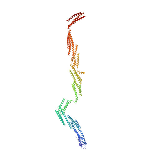
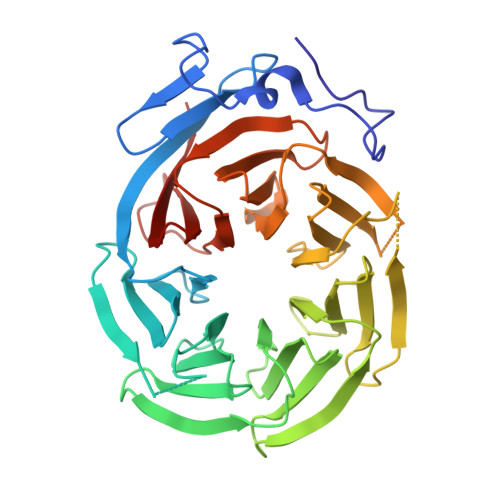
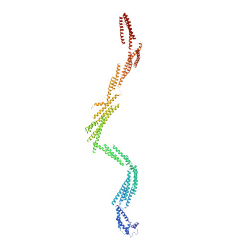
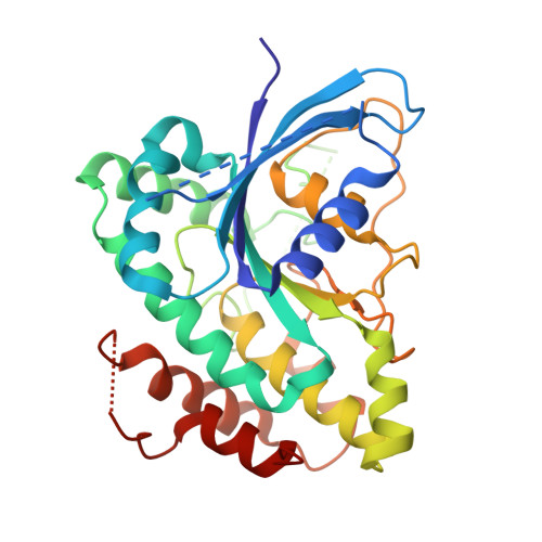
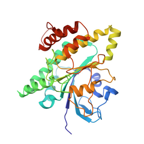
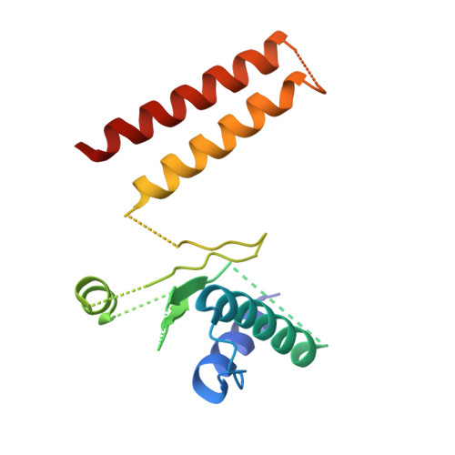
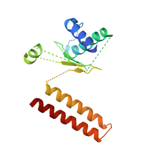
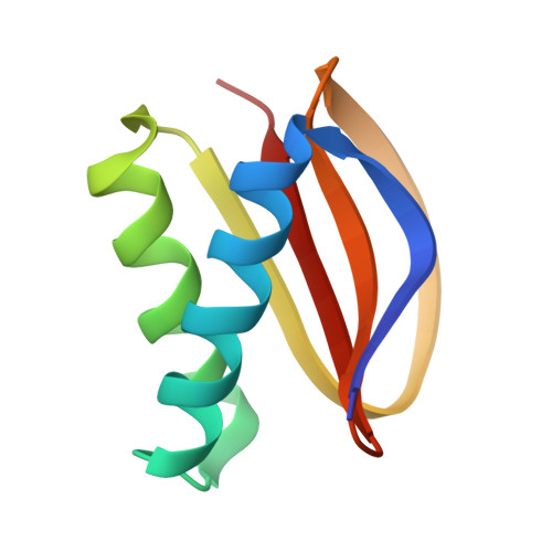
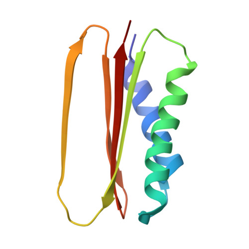
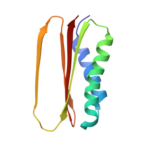
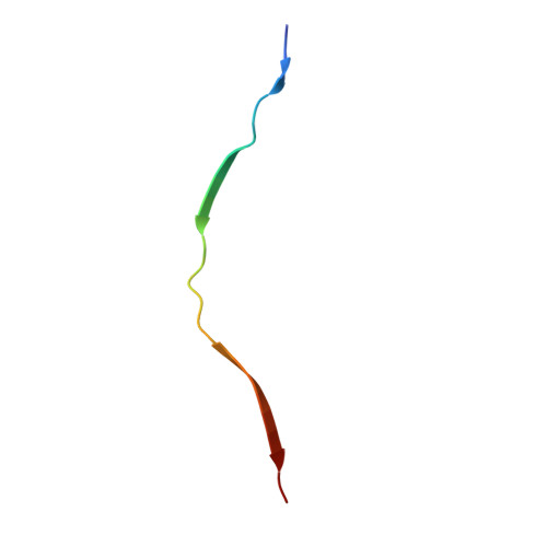
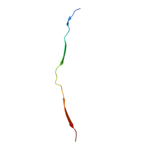
Cryo-EM Reveals How Human Cytoplasmic Dynein Is Auto-inhibited and Activated.

Zhang, K.Foster, H.E.Rondelet, A.Lacey, S.E.Bahi-Buisson, N.Bird, A.W.Carter, A.P.

(2017) Cell 169: 1303-1314.e18

  • DOI: https://doi.org/10.1016/j.cell.2017.05.025
  • Primary Citation Related Structures: 
    5NUG, 5NVS, 5NVU, 5NW4

  • PubMed Abstract: 

    Cytoplasmic dynein-1 binds dynactin and cargo adaptor proteins to form a transport machine capable of long-distance processive movement along microtubules. However, it is unclear why dynein-1 moves poorly on its own or how it is activated by dynactin. Here, we present a cryoelectron microscopy structure of the complete 1.4-megadalton human dynein-1 complex in an inhibited state known as the phi-particle. We reveal the 3D structure of the cargo binding dynein tail and show how self-dimerization of the motor domains locks them in a conformation with low microtubule affinity. Disrupting motor dimerization with structure-based mutagenesis drives dynein-1 into an open form with higher affinity for both microtubules and dynactin. We find the open form is also inhibited for movement and that dynactin relieves this by reorienting the motor domains to interact correctly with microtubules. Our model explains how dynactin binding to the dynein-1 tail directly stimulates its motor activity.


  • Organizational Affiliation
    • MRC Laboratory of Molecular Biology, Cambridge CB2 0QH, UK.

Macromolecule Content 

  • Total Structure Weight: 344.11 kDa 
  • Atom Count: 20,072 
  • Modeled Residue Count: 4,040 
  • Deposited Residue Count: 4,040 
  • Unique protein chains: 13

Macromolecules

Find similar proteins by:|  3D Structure
Entity ID: 1
MoleculeChains  Sequence LengthOrganismDetailsImage
dynein heavy chain932Homo sapiensMutation(s): 0 
Find similar proteins by:|  3D Structure
Entity ID: 2
MoleculeChains  Sequence LengthOrganismDetailsImage
dynein intermediate chainB [auth D],
C
350Homo sapiensMutation(s): 0 
Find similar proteins by:|  3D Structure
Entity ID: 3
MoleculeChains  Sequence LengthOrganismDetailsImage
dynein heavy chainD [auth B]893Homo sapiensMutation(s): 0 
Find similar proteins by:|  3D Structure
Entity ID: 4
MoleculeChains  Sequence LengthOrganismDetailsImage
dynein light intermediate chainE [auth F]298Homo sapiensMutation(s): 0 
Find similar proteins by:|  3D Structure
Entity ID: 5
MoleculeChains  Sequence LengthOrganismDetailsImage
dynein light intermediate chainF [auth E]295Homo sapiensMutation(s): 0 
Find similar proteins by:|  3D Structure
Entity ID: 6
MoleculeChains  Sequence LengthOrganismDetailsImage
N-terminal dimerization domainG [auth 2]125Homo sapiensMutation(s): 0 
Find similar proteins by:|  3D Structure
Entity ID: 7
MoleculeChains  Sequence LengthOrganismDetailsImage
N-terminal dimerization domainH [auth 1]124Homo sapiensMutation(s): 0 
Find similar proteins by:|  3D Structure
Entity ID: 8
MoleculeChains  Sequence LengthOrganismDetailsImage
LC8
I, J
85Homo sapiensMutation(s): 0 
Find similar proteins by:|  3D Structure
Entity ID: 9
MoleculeChains  Sequence LengthOrganismDetailsImage
Tctex103Homo sapiensMutation(s): 0 
Find similar proteins by:|  3D Structure
Entity ID: 10
MoleculeChains  Sequence LengthOrganismDetailsImage
Tctex104Homo sapiensMutation(s): 0 
Find similar proteins by:|  3D Structure
Entity ID: 11
MoleculeChains  Sequence LengthOrganismDetailsImage
intermediate chain27Homo sapiensMutation(s): 0 
Find similar proteins by:|  3D Structure
Entity ID: 12
MoleculeChains  Sequence LengthOrganismDetailsImage
intermediate chain29Homo sapiensMutation(s): 0 
Find similar proteins by:|  3D Structure
Entity ID: 13
MoleculeChains  Sequence LengthOrganismDetailsImage
RoblO [auth R],
P [auth S]
120Homo sapiensMutation(s): 0 

Experimental Data & Validation

Experimental Data

  • Method: ELECTRON MICROSCOPY
  • Resolution: 8.40 Å
  • Aggregation State: PARTICLE 
  • Reconstruction Method: SINGLE PARTICLE 
EM Software:
TaskSoftware PackageVersion
RECONSTRUCTIONRELION1.4

Structure Validation

View Full Validation Report


Entry History 

& Funding Information

Deposition Data


Funding OrganizationLocationGrant Number
Wellcome TrustUnited KingdomWT100387
Medical Research Council (United Kingdom)United KingdomMC_UP_A025_1011

Revision History  (Full details and data files)

  • Version 1.0: 2017-06-28
    Type: Initial release
  • Version 1.1: 2017-08-30
    Changes: Author supporting evidence, Data collection
  • Version 1.2: 2019-02-06
    Changes: Advisory, Data collection, Derived calculations
  • Version 1.3: 2024-05-15
    Changes: Database references
  • Version 1.4: 2024-07-10
    Changes: Data collection