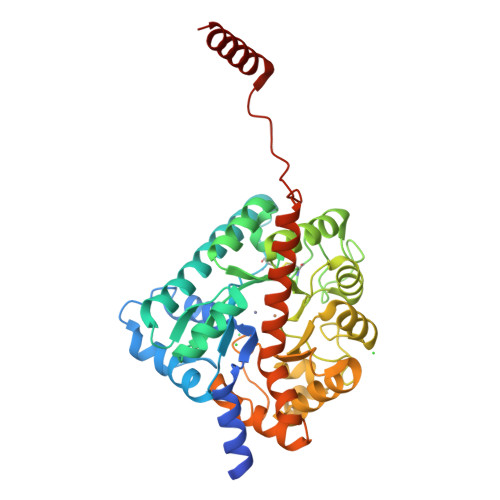
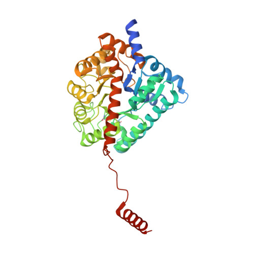
5NS5

Cys-Gly dipeptidase GliJ in complex with Cu2+ and Zn2+


Experimental Data Snapshot

  • Method: X-RAY DIFFRACTION
  • Resolution: 2.20 Å
  • R-Value Free: 0.189 
  • R-Value Work: 0.177 
  • R-Value Observed: 0.177 

Starting Model: experimental
View more details

wwPDB Validation   3D Report Full Report


This is version 1.3 of the entry. See complete history


Literature

Gliotoxin Biosynthesis: Structure, Mechanism, and Metal Promiscuity of Carboxypeptidase GliJ.

Marion, A.Groll, M.Scharf, D.H.Scherlach, K.Glaser, M.Sievers, H.Schuster, M.Hertweck, C.Brakhage, A.A.Antes, I.Huber, E.M.

(2017) ACS Chem Biol 12: 1874-1882

  • DOI: https://doi.org/10.1021/acschembio.6b00847
  • Primary Citation of Related Structures:  
    5LWZ, 5LX0, 5LX1, 5LX4, 5LX7, 5NRT, 5NRU, 5NRX, 5NRY, 5NRZ, 5NS1, 5NS2, 5NS5

  • PubMed Abstract: 

    The formation of glutathione (GSH) conjugates, best known from the detoxification of xenobiotics, is a widespread strategy to incorporate sulfur into biomolecules. The biosynthesis of gliotoxin, a virulence factor of the human pathogenic fungus Aspergillus fumigatus, involves attachment of two GSH molecules and their sequential decomposition to yield two reactive thiol groups. The degradation of the GSH moieties requires the activity of the Cys-Gly carboxypeptidase GliJ, for which we describe the X-ray structure here. The enzyme forms a homodimer with each monomer comprising one active site. Two metal ions are present per proteolytic center, thus assigning GliJ to the diverse family of dinuclear metallohydrolases. Depending on availability, Zn 2+ , Fe 2+ , Fe 3+ , Mn 2+ , Cu 2+ , Co 2+ , or Ni 2+ ions are accepted as cofactors. Despite this high metal promiscuity, a preference for zinc versus iron and manganese was noted. Mutagenesis experiments revealed details of metal coordination, and molecular modeling delivered insights into substrate recognition and processing by GliJ. The latter results suggest a reaction mechanism in which the two scissile peptide bonds of one gliotoxin precursor molecule are hydrolyzed sequentially and in a given order.


  • Organizational Affiliation

    Center for Integrated Protein Science Munich at the Department of Biosciences, Technische Universität München , Emil-Erlenmeyer-Forum 8, D-85354 Freising, Germany.


Macromolecules
Find similar proteins by:  (by identity cutoff)  |  3D Structure
Entity ID: 1
MoleculeChains Sequence LengthOrganismDetailsImage
Dipeptidase gliJ401Aspergillus fumigatusMutation(s): 0 
Gene Names: gliJAFUA_6G09650
EC: 3.4.13.19
UniProt
Find proteins for Q4WMJ8 (Aspergillus fumigatus (strain ATCC MYA-4609 / CBS 101355 / FGSC A1100 / Af293))
Explore Q4WMJ8 
Go to UniProtKB:  Q4WMJ8
Entity Groups  
Sequence Clusters30% Identity50% Identity70% Identity90% Identity95% Identity100% Identity
UniProt GroupQ4WMJ8
Sequence Annotations
Expand
  • Reference Sequence
Small Molecules
Ligands 3 Unique
IDChains Name / Formula / InChI Key2D Diagram3D Interactions
ZN
Query on ZN

Download Ideal Coordinates CCD File 
B [auth A],
C [auth A]
ZINC ION
Zn
PTFCDOFLOPIGGS-UHFFFAOYSA-N
CU
Query on CU

Download Ideal Coordinates CCD File 
D [auth A],
E [auth A]
COPPER (II) ION
Cu
JPVYNHNXODAKFH-UHFFFAOYSA-N
CL
Query on CL

Download Ideal Coordinates CCD File 
F [auth A],
G [auth A],
H [auth A]
CHLORIDE ION
Cl
VEXZGXHMUGYJMC-UHFFFAOYSA-M
Experimental Data & Validation

Experimental Data

  • Method: X-RAY DIFFRACTION
  • Resolution: 2.20 Å
  • R-Value Free: 0.189 
  • R-Value Work: 0.177 
  • R-Value Observed: 0.177 
  • Space Group: P 32 2 1
Unit Cell:
Length ( Å )Angle ( ˚ )
a = 99α = 90
b = 99β = 90
c = 106.49γ = 120
Software Package:
Software NamePurpose
REFMACrefinement
XDSdata reduction
XSCALEdata scaling
REFMACphasing

Structure Validation

View Full Validation Report


Entry History & Funding Information

Deposition Data


Funding OrganizationLocationGrant Number
Hans-Fischer-GesellschaftGermany--
German Research FoundationGermanySFB749

Revision History  (Full details and data files)

  • Version 1.0: 2017-05-31
    Type: Initial release
  • Version 1.1: 2017-08-02
    Changes: Database references
  • Version 1.2: 2024-01-17
    Changes: Author supporting evidence, Data collection, Database references, Derived calculations, Refinement description
  • Version 1.3: 2024-10-16
    Changes: Structure summary