


Mtb TMK crystal structure in complex with compound LS3080
Merceron, R., Song, L., Munier-Lehmann, H., Van Calenbergh, S., Savvides, S.To be published.
Experimental Data Snapshot
Starting Model: experimental
View more details
Entity ID: 1 | |||||
|---|---|---|---|---|---|
| Molecule | Chains | Sequence Length | Organism | Details | Image |
| Thymidylate kinase | 214 | Mycobacterium tuberculosis H37Rv | Mutation(s): 0 Gene Names: tmk, Rv3247c EC: 2.7.4.9 | 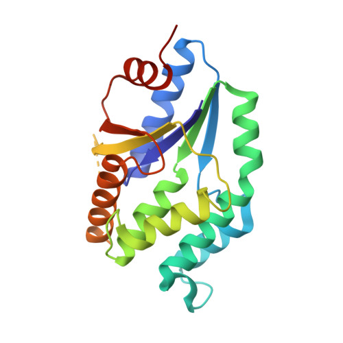 | |
UniProt | |||||
Entity Groups | |||||
| Sequence Clusters | 30% Identity50% Identity70% Identity90% Identity95% Identity100% Identity | ||||
| UniProt Group | P9WKE1 | ||||
Sequence AnnotationsExpand | |||||
Reference Sequence | |||||
| Ligands 2 Unique | |||||
|---|---|---|---|---|---|
| ID | Chains | Name / Formula / InChI Key | 2D Diagram | 3D Interactions | |
| 95W Download:Ideal Coordinates CCD File | C [auth A], G [auth B] | 5-methyl-1-[1-[(6-phenoxypyridin-2-yl)methyl]piperidin-4-yl]pyrimidine-2,4-dione C22 H24 N4 O3 BKRRWLAIGUKKJX-UHFFFAOYSA-N |  | ||
| CL Download:Ideal Coordinates CCD File | D [auth A] E [auth A] F [auth A] H [auth B] I [auth B] | CHLORIDE ION Cl VEXZGXHMUGYJMC-UHFFFAOYSA-M |  | ||
| Length ( Å ) | Angle ( ˚ ) |
|---|---|
| a = 130.23 | α = 90 |
| b = 130.23 | β = 90 |
| c = 120.19 | γ = 90 |
| Software Name | Purpose |
|---|---|
| PHENIX | refinement |
| XDS | data reduction |
| XSCALE | data scaling |
| MOLREP | phasing |


