5NQB | pdb_00005nqb

Rabbit Muscle L-lactate dehydrogenase in complex with malonate


Experimental Data Snapshot

  • Method: X-RAY DIFFRACTION
  • Resolution: 1.58 Å
  • R-Value Free: 
    0.218 (Depositor), 0.217 (DCC) 
  • R-Value Work: 
    0.193 (Depositor), 0.192 (DCC) 
  • R-Value Observed: 
    0.194 (Depositor) 

Starting Model: experimental
View more details

wwPDB Validation 3D Report Full Report

Validation slider image for 5NQB

This is version 1.3 of the entry. See complete history

Literature

The self-inhibitory nature of metabolic networks and its alleviation through compartmentalization.

Alam, M.T.Olin-Sandoval, V.Stincone, A.Keller, M.A.Zelezniak, A.Luisi, B.F.Ralser, M.

(2017) Nat Commun 8: 16018-16018

  • DOI: https://doi.org/10.1038/ncomms16018
  • Primary Citation Related Structures: 
    5NQB, 5NQQ

  • PubMed Abstract: 

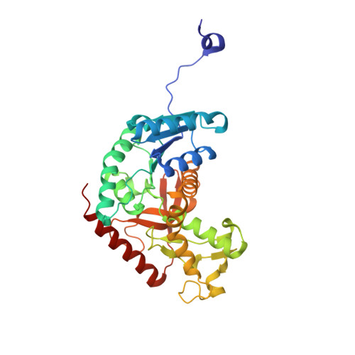
    Metabolites can inhibit the enzymes that generate them. To explore the general nature of metabolic self-inhibition, we surveyed enzymological data accrued from a century of experimentation and generated a genome-scale enzyme-inhibition network. Enzyme inhibition is often driven by essential metabolites, affects the majority of biochemical processes, and is executed by a structured network whose topological organization is reflecting chemical similarities that exist between metabolites. Most inhibitory interactions are competitive, emerge in the close neighbourhood of the inhibited enzymes, and result from structural similarities between substrate and inhibitors. Structural constraints also explain one-third of allosteric inhibitors, a finding rationalized by crystallographic analysis of allosterically inhibited L-lactate dehydrogenase. Our findings suggest that the primary cause of metabolic enzyme inhibition is not the evolution of regulatory metabolite-enzyme interactions, but a finite structural diversity prevalent within the metabolome. In eukaryotes, compartmentalization minimizes inevitable enzyme inhibition and alleviates constraints that self-inhibition places on metabolism.


  • Organizational Affiliation
    • Department of Biochemistry and Cambridge Systems Biology Centre, University of Cambridge, 80 Tennis Court Road, Cambridge CB2 1GA, UK.

Macromolecule Content 

  • Total Structure Weight: 147.11 kDa 
  • Atom Count: 11,632 
  • Modeled Residue Count: 1,324 
  • Deposited Residue Count: 1,328 
  • Unique protein chains: 1

Macromolecules

Find similar proteins by:|  3D Structure
Entity ID: 1
MoleculeChains  Sequence LengthOrganismDetailsImage
L-lactate dehydrogenase A chain
A, B, C, D
332Oryctolagus cuniculusMutation(s): 1 
Gene Names: LDHA
EC: 1.1.1.27
UniProt
Find proteins for P13491 (Oryctolagus cuniculus)
Explore P13491 
Go to UniProtKB:  P13491
Entity Groups
Sequence Clusters30% Identity50% Identity70% Identity90% Identity95% Identity100% Identity
UniProt GroupP13491
Sequence Annotations
Expand
Reference Sequence

Experimental Data & Validation

Experimental Data

  • Method: X-RAY DIFFRACTION
  • Resolution: 1.58 Å
  • R-Value Free:  0.218 (Depositor), 0.217 (DCC) 
  • R-Value Work:  0.193 (Depositor), 0.192 (DCC) 
  • R-Value Observed: 0.194 (Depositor) 
Space Group: P 1 21 1
Unit Cell:
Length ( Å )Angle ( ˚ )
a = 70.806α = 90
b = 76.952β = 96.48
c = 118.858γ = 90
Software Package:
Software NamePurpose
PHENIXrefinement
MOSFLMdata reduction
Aimlessdata scaling
PHASERphasing

Structure Validation

View Full Validation Report


Entry History 

& Funding Information

Deposition Data


Funding OrganizationLocationGrant Number
Wellcome TrustUnited Kingdom094229/Z/10/Z

Revision History  (Full details and data files)

  • Version 1.0: 2017-05-03
    Type: Initial release
  • Version 1.1: 2017-07-19
    Changes: Database references
  • Version 1.2: 2019-10-16
    Changes: Data collection
  • Version 1.3: 2024-01-17
    Changes: Data collection, Database references, Refinement description