5NQA | pdb_00005nqa

Crystal structure of GalNAc-T4 in complex with the monoglycopeptide 3


Experimental Data Snapshot

  • Method: X-RAY DIFFRACTION
  • Resolution: 1.90 Å
  • R-Value Free: 
    0.274 (Depositor), 0.280 (DCC) 
  • R-Value Work: 
    0.225 (Depositor), 0.229 (DCC) 
  • R-Value Observed: 
    0.226 (Depositor) 

Starting Model: experimental
View more details

wwPDB Validation 3D Report Full Report

Validation slider image for 5NQA

Ligand Structure Quality Assessment 


This is version 1.3 of the entry. See complete history

Literature

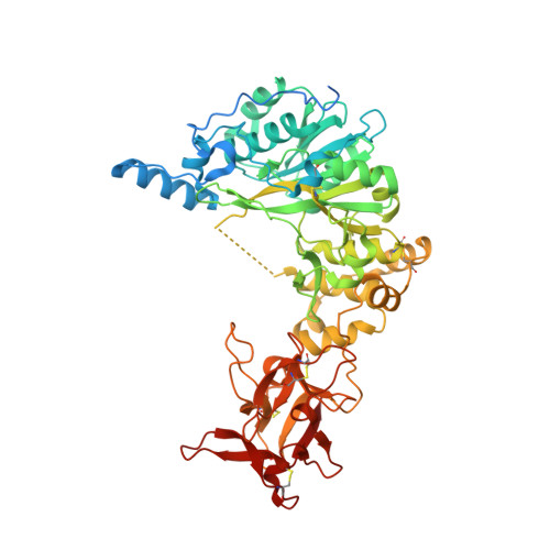
The interdomain flexible linker of the polypeptide GalNAc transferases dictates their long-range glycosylation preferences.

Rivas, M.L.Lira-Navarrete, E.Daniel, E.J.P.Companon, I.Coelho, H.Diniz, A.Jimenez-Barbero, J.Peregrina, J.M.Clausen, H.Corzana, F.Marcelo, F.Jimenez-Oses, G.Gerken, T.A.Hurtado-Guerrero, R.

(2017) Nat Commun 8: 1959-1959

  • DOI: https://doi.org/10.1038/s41467-017-02006-0
  • Primary Citation Related Structures: 
    5NQA

  • PubMed Abstract: 

    The polypeptide GalNAc-transferases (GalNAc-Ts), that initiate mucin-type O-glycosylation, consist of a catalytic and a lectin domain connected by a flexible linker. In addition to recognizing polypeptide sequence, the GalNAc-Ts exhibit unique long-range N- and/or C-terminal prior glycosylation (GalNAc-O-Ser/Thr) preferences modulated by the lectin domain. Here we report studies on GalNAc-T4 that reveal the origins of its unique N-terminal long-range glycopeptide specificity, which is the opposite of GalNAc-T2. The GalNAc-T4 structure bound to a monoglycopeptide shows that the GalNAc-binding site of its lectin domain is rotated relative to the homologous GalNAc-T2 structure, explaining their different long-range preferences. Kinetics and molecular dynamics simulations on several GalNAc-T2 flexible linker constructs show altered remote prior glycosylation preferences, confirming that the flexible linker dictates the rotation of the lectin domain, thus modulating the GalNAc-Ts' long-range preferences. This work for the first time provides the structural basis for the different remote prior glycosylation preferences of the GalNAc-Ts.


  • Organizational Affiliation
    • BIFI, University of Zaragoza, BIFI-IQFR (CSIC) Joint Unit, Mariano Esquillor s/n, Campus Rio Ebro, Edificio I+D, Zaragoza, 50018, Spain.

Macromolecule Content 

  • Total Structure Weight: 140.48 kDa 
  • Atom Count: 10,430 
  • Modeled Residue Count: 1,029 
  • Deposited Residue Count: 1,172 
  • Unique protein chains: 2

Macromolecules

Find similar proteins by:|  3D Structure
Entity ID: 1
MoleculeChains  Sequence LengthOrganismDetailsImage
Polypeptide N-acetylgalactosaminyltransferase 4
A, B
578Homo sapiensMutation(s): 0 
Gene Names: GALNT4
EC: 2.4.1.41
UniProt & NIH Common Fund Data Resources
Find proteins for Q8N4A0 (Homo sapiens)
Explore Q8N4A0 
Go to UniProtKB:  Q8N4A0
PHAROS:  Q8N4A0
GTEx:  ENSG00000257594 
Entity Groups
Sequence Clusters30% Identity50% Identity70% Identity90% Identity95% Identity100% Identity
UniProt GroupQ8N4A0
Sequence Annotations
Expand
Reference Sequence
Find similar proteins by:  Sequence   |   3D Structure  
Entity ID: 2
MoleculeChains  Sequence LengthOrganismDetailsImage
Monoglycopeptide 3C [auth G]16Homo sapiensMutation(s): 0 

Small Molecules

Ligands 3 Unique
IDChains Name / Formula / InChI Key2D Diagram3D Interactions
NGA

Query on NGA


Download:Ideal Coordinates CCD File
HC [auth G],
UA [auth A]
2-acetamido-2-deoxy-beta-D-galactopyranose
C8 H15 N O6
OVRNDRQMDRJTHS-JAJWTYFOSA-N
GOL

Query on GOL


Download:Ideal Coordinates CCD File
DC [auth B]
EC [auth B]
FC [auth B]
GC [auth B]
OA [auth A]
DC [auth B],
EC [auth B],
FC [auth B],
GC [auth B],
OA [auth A],
PA [auth A],
QA [auth A],
RA [auth A],
SA [auth A],
TA [auth A]
GLYCEROL
C3 H8 O3
PEDCQBHIVMGVHV-UHFFFAOYSA-N
EDO

Query on EDO


Download:Ideal Coordinates CCD File
AA [auth A]
AB [auth B]
AC [auth B]
BA [auth A]
BB [auth B]
AA [auth A],
AB [auth B],
AC [auth B],
BA [auth A],
BB [auth B],
BC [auth B],
CA [auth A],
CB [auth B],
CC [auth B],
D [auth A],
DA [auth A],
DB [auth B],
E [auth A],
EA [auth A],
EB [auth B],
F [auth A],
FA [auth A],
FB [auth B],
G [auth A],
GA [auth A],
GB [auth B],
H [auth A],
HA [auth A],
HB [auth B],
I [auth A],
IA [auth A],
IB [auth B],
J [auth A],
JA [auth A],
JB [auth B],
K [auth A],
KA [auth A],
KB [auth B],
L [auth A],
LA [auth A],
LB [auth B],
M [auth A],
MA [auth A],
MB [auth B],
N [auth A],
NA [auth A],
NB [auth B],
O [auth A],
OB [auth B],
P [auth A],
PB [auth B],
Q [auth A],
QB [auth B],
R [auth A],
RB [auth B],
S [auth A],
SB [auth B],
T [auth A],
TB [auth B],
U [auth A],
UB [auth B],
V [auth A],
VA [auth B],
VB [auth B],
W [auth A],
WA [auth B],
WB [auth B],
X [auth A],
XA [auth B],
XB [auth B],
Y [auth A],
YA [auth B],
YB [auth B],
Z [auth A],
ZA [auth B],
ZB [auth B]
1,2-ETHANEDIOL
C2 H6 O2
LYCAIKOWRPUZTN-UHFFFAOYSA-N

Experimental Data & Validation

Experimental Data

  • Method: X-RAY DIFFRACTION
  • Resolution: 1.90 Å
  • R-Value Free:  0.274 (Depositor), 0.280 (DCC) 
  • R-Value Work:  0.225 (Depositor), 0.229 (DCC) 
  • R-Value Observed: 0.226 (Depositor) 
Space Group: P 1
Unit Cell:
Length ( Å )Angle ( ˚ )
a = 65.447α = 116.47
b = 79.881β = 96.74
c = 88.614γ = 104.72
Software Package:
Software NamePurpose
REFMACrefinement
XDSdata reduction
SCALAdata scaling
PHASERphasing

Structure Validation

View Full Validation Report


Ligand Structure Quality Assessment 


Entry History 

Revision History  (Full details and data files)

  • Version 1.0: 2017-12-20
    Type: Initial release
  • Version 1.1: 2020-07-29
    Type: Remediation
    Reason: Carbohydrate remediation
    Changes: Data collection, Derived calculations, Structure summary
  • Version 1.2: 2024-01-17
    Changes: Data collection, Database references, Refinement description, Structure summary
  • Version 1.3: 2024-10-16
    Changes: Structure summary