5NOQ

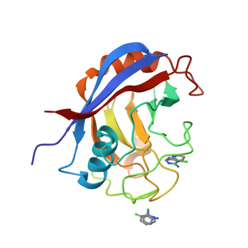
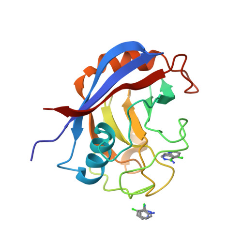
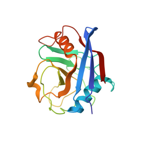
Structure of cyclophilin A in complex with 3-chloropyridin-2-amine


Experimental Data Snapshot

  • Method: X-RAY DIFFRACTION
  • Resolution: 1.60 Å
  • R-Value Free: 0.196 
  • R-Value Work: 0.165 
  • R-Value Observed: 0.167 

Starting Model: experimental
View more details

wwPDB Validation   3D Report Full Report


This is version 1.2 of the entry. See complete history


Literature

Pushing the Limits of Detection of Weak Binding Using Fragment-Based Drug Discovery: Identification of New Cyclophilin Binders.

Georgiou, C.McNae, I.Wear, M.Ioannidis, H.Michel, J.Walkinshaw, M.

(2017) J Mol Biol 429: 2556-2570

  • DOI: https://doi.org/10.1016/j.jmb.2017.06.016
  • Primary Citation of Related Structures:  
    5NOQ, 5NOR, 5NOS, 5NOT, 5NOU, 5NOV, 5NOW, 5NOX, 5NOY, 5NOZ

  • PubMed Abstract: 

    Fragment-based drug discovery is an increasingly popular method to identify novel small-molecule drug candidates. One of the limitations of the approach is the difficulty of accurately characterizing weak binding events. This work reports a combination of X-ray diffraction, surface plasmon resonance experiments and molecular dynamics simulations for the characterization of binders to different isoforms of the cyclophilin (Cyp) protein family. Although several Cyp inhibitors have been reported in the literature, it has proven challenging to achieve high binding selectivity for different isoforms of this protein family. The present studies have led to the identification of several structurally novel fragments that bind to diverse Cyp isoforms in distinct pockets with low millimolar dissociation constants. A detailed comparison of the merits and drawbacks of the experimental and computational techniques is presented, and emerging strategies for designing ligands with enhanced isoform specificity are described.


  • Organizational Affiliation

    University of Edinburgh, Michael Swann Building, Max Born Crescent, Edinburgh, Scotland, EH9 3BF, UK; University of Edinburgh, Joseph Black Building, David Brewster Road, Edinburgh, Scotland, EH9 3FJ, UK.


Macromolecules
Find similar proteins by:  (by identity cutoff)  |  3D Structure
Entity ID: 1
MoleculeChains Sequence LengthOrganismDetailsImage
Peptidyl-prolyl cis-trans isomerase A165Homo sapiensMutation(s): 0 
Gene Names: PPIACYPA
EC: 5.2.1.8
UniProt & NIH Common Fund Data Resources
Find proteins for P62937 (Homo sapiens)
Explore P62937 
Go to UniProtKB:  P62937
PHAROS:  P62937
GTEx:  ENSG00000196262 
Entity Groups  
Sequence Clusters30% Identity50% Identity70% Identity90% Identity95% Identity100% Identity
UniProt GroupP62937
Sequence Annotations
Expand
  • Reference Sequence
Experimental Data & Validation

Experimental Data

  • Method: X-RAY DIFFRACTION
  • Resolution: 1.60 Å
  • R-Value Free: 0.196 
  • R-Value Work: 0.165 
  • R-Value Observed: 0.167 
  • Space Group: P 21 21 21
Unit Cell:
Length ( Å )Angle ( ˚ )
a = 42.18α = 90
b = 53.88β = 90
c = 85.91γ = 90
Software Package:
Software NamePurpose
REFMACrefinement
SCALAdata scaling
PDB_EXTRACTdata extraction
MOSFLMdata reduction
PHASERphasing

Structure Validation

View Full Validation Report


Entry History & Funding Information

Deposition Data


Funding OrganizationLocationGrant Number
Biotechnology and Biological Sciences Research CouncilUnited Kingdom--

Revision History  (Full details and data files)

  • Version 1.0: 2017-07-12
    Type: Initial release
  • Version 1.1: 2017-08-16
    Changes: Database references
  • Version 1.2: 2024-01-17
    Changes: Data collection, Database references, Refinement description