5NOP | pdb_00005nop

Structure of Mojiang virus attachment glycoprotein


Experimental Data Snapshot

  • Method: X-RAY DIFFRACTION
  • Resolution: 1.94 Å
  • R-Value Free: 
    0.222 (Depositor), 0.223 (DCC) 
  • R-Value Work: 
    0.182 (Depositor), 0.182 (DCC) 
  • R-Value Observed: 
    0.184 (Depositor) 

Starting Model: experimental
View more details

wwPDB Validation 3D Report Full Report

Validation slider image for 5NOP

This is version 1.4 of the entry. See complete history

Literature

Idiosyncratic Mojiang virus attachment glycoprotein directs a host-cell entry pathway distinct from genetically related henipaviruses.

Rissanen, I.Ahmed, A.A.Azarm, K.Beaty, S.Hong, P.Nambulli, S.Duprex, W.P.Lee, B.Bowden, T.A.

(2017) Nat Commun 8: 16060-16060

  • DOI: https://doi.org/10.1038/ncomms16060
  • Primary Citation Related Structures: 
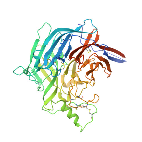
    5NOP

  • PubMed Abstract: 

    In 2012, cases of lethal pneumonia among Chinese miners prompted the isolation of a rat-borne henipavirus (HNV), Mòjiāng virus (MojV). Although MojV is genetically related to highly pathogenic bat-borne henipaviruses, the absence of a conserved ephrin receptor-binding motif in the MojV attachment glycoprotein (MojV-G) indicates a differing host-cell recognition mechanism. Here we find that MojV-G displays a six-bladed β-propeller fold bearing limited similarity to known paramyxoviral attachment glycoproteins, in particular at host receptor-binding surfaces. We confirm the inability of MojV-G to interact with known paramyxoviral receptors in vitro, indicating an independence from well-characterized ephrinB2/B3, sialic acid and CD150-mediated entry pathways. Furthermore, we find that MojV-G is antigenically distinct, indicating that MojV would less likely be detected in existing large-scale serological screening studies focused on well-established HNVs. Altogether, these data indicate a unique host-cell entry pathway for this emerging and potentially pathogenic HNV.


  • Organizational Affiliation
    • Division of Structural Biology, Wellcome Trust Centre for Human Genetics, University of Oxford, Roosevelt Drive, Oxford, Oxfordshire OX3 7BN, UK.

Macromolecule Content 

  • Total Structure Weight: 104.9 kDa 
  • Atom Count: 7,629 
  • Modeled Residue Count: 857 
  • Deposited Residue Count: 942 
  • Unique protein chains: 1

Macromolecules

Find similar proteins by:|  3D Structure
Entity ID: 1
MoleculeChains  Sequence LengthOrganismDetailsImage
Attachment glycoprotein
A, B
471Mojiang virusMutation(s): 0 
Gene Names: GMojVGP5
UniProt
Find proteins for W8TIR7 (Mojiang virus)
Explore W8TIR7 
Go to UniProtKB:  W8TIR7
Entity Groups
Sequence Clusters30% Identity50% Identity70% Identity90% Identity95% Identity100% Identity
UniProt GroupW8TIR7
Sequence Annotations
Expand
Reference Sequence

Experimental Data & Validation

Experimental Data

  • Method: X-RAY DIFFRACTION
  • Resolution: 1.94 Å
  • R-Value Free:  0.222 (Depositor), 0.223 (DCC) 
  • R-Value Work:  0.182 (Depositor), 0.182 (DCC) 
  • R-Value Observed: 0.184 (Depositor) 
Space Group: P 21 21 21
Unit Cell:
Length ( Å )Angle ( ˚ )
a = 60.04α = 90
b = 102.31β = 90
c = 162.06γ = 90
Software Package:
Software NamePurpose
xia2data reduction
xia2data scaling
PHENIXphasing
PHENIXrefinement
PDB_EXTRACTdata extraction

Structure Validation

View Full Validation Report


Entry History 

& Funding Information

Deposition Data


Funding OrganizationLocationGrant Number
Medical Research Council (United Kingdom)United KingdomMR/L009528/1

Revision History  (Full details and data files)

  • Version 1.0: 2017-07-19
    Type: Initial release
  • Version 1.1: 2017-07-26
    Changes: Database references
  • Version 1.2: 2019-04-03
    Changes: Data collection, Source and taxonomy
  • Version 1.3: 2024-01-17
    Changes: Data collection, Database references, Refinement description
  • Version 1.4: 2024-11-13
    Changes: Structure summary