5NI9 | pdb_00005ni9

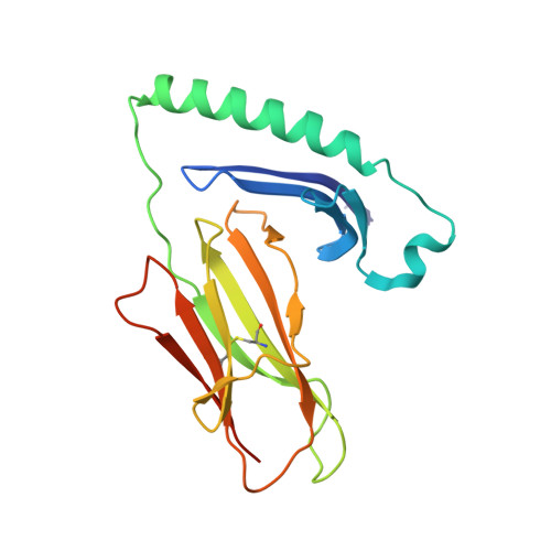
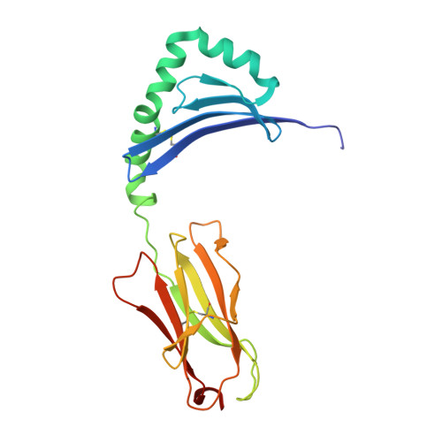
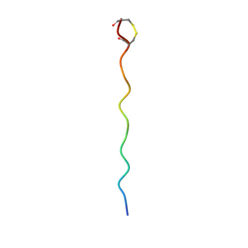
Crystal structure of HLA-DRB1*04:01 with the alpha-enolase peptide 326-340


Experimental Data Snapshot

  • Method: X-RAY DIFFRACTION
  • Resolution: 1.33 Å
  • R-Value Free: 
    0.176 (Depositor), 0.176 (DCC) 
  • R-Value Work: 
    0.146 (Depositor), 0.146 (DCC) 
  • R-Value Observed: 
    0.146 (Depositor) 

Starting Model: experimental
View more details

wwPDB Validation 3D Report Full Report

Validation slider image for 5NI9

This is version 1.5 of the entry. See complete history. 

Literature

Memory T cells specific to citrullinated alpha-enolase are enriched in the rheumatic joint

Pieper, J., Dubnovitsky, A., Gerstner, C., James, E.A., Rieck, M., Kozhukh, G., Tandre, K., Pellegrino, S., Gebe, J.A., Ronnblom, L., Sandalova, T., Kwok, W.W., Klareskog, L., Buckner, J.H., Achour, A., Malmstrom, V.

(2018) J Autoimmun 92: 47-56

  • DOI: https://doi.org/10.1016/j.jaut.2018.04.004
  • Primary Citation Related Structures: 
    5NI9, 5NIG

  • PubMed Abstract: 

    ACPA-positive rheumatoid arthritis (RA) is associated with distinct HLA-DR alleles and immune responses to many citrullinated self-antigens. Herein we investigated the T cell epitope confined within α-enolase 326-340 in the context of HLA-DRB1*04:01 and assessed the corresponding CD4 + T cells in both the circulation and in the rheumatic joint. Comparative crystallographic analyses were performed for the native and citrullinated α-enolase 326-340 peptides in complex with HLA-DRB1*04:01. HLA-tetramers assembled with either the native or citrullinated peptide were used for ex vivo and in vitro assessment of α-enolase-specific T cells in peripheral blood, synovial fluid and synovial tissue by flow cytometry. The native and modified peptides take a completely conserved structural conformation within the peptide-binding cleft of HLA-DRB1*04:01. The citrulline residue-327 was located N-terminally, protruding towards TCRs. The frequencies of T cells recognizing native eno 326-340 were similar in synovial fluid and peripheral blood, while in contrast, the frequency of T cells recognizing cit-eno 326-340 was significantly elevated in synovial fluid compared to peripheral blood (3.6-fold, p = 0.0150). Additionally, citrulline-specific T cells with a memory phenotype were also significantly increased (1.6-fold, p = 0.0052) in synovial fluid compared to peripheral blood. The native T cell epitope confined within α-enolase 326-340 does not appear to lead to complete negative selection of cognate CD4 + T cells. In RA patient samples, only T cells recognizing the citrullinated version of α-enolase 326-340 were found at elevated frequencies implicating that neo-antigen formation is critical for breach of tolerance.


  • Organizational Affiliation: 
    • Rheumatology Unit, Department of Medicine Solna, Center for Molecular Medicine, Karolinska Institutet, Karolinska University Hospital, Stockholm, Sweden.

Macromolecule Content 

  • Total Structure Weight: 47.17 kDa 
  • Atom Count: 3,606 
  • Modeled Residue Count: 386 
  • Deposited Residue Count: 402 
  • Unique protein chains: 3

Macromolecules

Find similar proteins by:|  3D Structure
Entity ID: 1
MoleculeChains  Sequence LengthOrganismDetailsImage
HLA class II histocompatibility antigen, DR alpha chain189Homo sapiensMutation(s): 0 
Gene Names: HLA-DRA, HLA-DRA1
UniProt & NIH Common Fund Data Resources
Find proteins for P01903 (Homo sapiens)
Explore P01903 
Go to UniProtKB:  P01903
PHAROS:  P01903
GTEx:  ENSG00000204287 
Entity Groups
Sequence Clusters30% Identity50% Identity70% Identity90% Identity95% Identity100% Identity
UniProt GroupP01903
Sequence Annotations
Expand
Reference Sequence
Find similar proteins by:|  3D Structure
Entity ID: 2
MoleculeChains  Sequence LengthOrganismDetailsImage
HLA class II histocompatibility antigen, DRB1-4 beta chain198Homo sapiensMutation(s): 0 
Gene Names: HLA-DRB1
UniProt & NIH Common Fund Data Resources
Find proteins for P01911 (Homo sapiens)
Explore P01911 
Go to UniProtKB:  P01911
PHAROS:  P01911
GTEx:  ENSG00000196126 
Entity Groups
Sequence Clusters30% Identity50% Identity70% Identity90% Identity95% Identity100% Identity
UniProt GroupP01911
Sequence Annotations
Expand
Reference Sequence
Find similar proteins by:  Sequence   |   3D Structure  
Entity ID: 3
MoleculeChains  Sequence LengthOrganismDetailsImage
Alpha-enolase15Homo sapiensMutation(s): 0 
EC: 4.2.1.11
UniProt & NIH Common Fund Data Resources
Find proteins for P06733 (Homo sapiens)
Explore P06733 
Go to UniProtKB:  P06733
PHAROS:  P06733
GTEx:  ENSG00000074800 
Entity Groups
Sequence Clusters30% Identity50% Identity70% Identity90% Identity95% Identity100% Identity
UniProt GroupP06733
Sequence Annotations
Expand
Reference Sequence

Experimental Data & Validation

Experimental Data

  • Method: X-RAY DIFFRACTION
  • Resolution: 1.33 Å
  • R-Value Free:  0.176 (Depositor), 0.176 (DCC) 
  • R-Value Work:  0.146 (Depositor), 0.146 (DCC) 
  • R-Value Observed: 0.146 (Depositor) 
Space Group: P 21 21 2
Unit Cell:
Length ( Å )Angle ( ˚ )
a = 68.447α = 90
b = 128.109β = 90
c = 53.585γ = 90
Software Package:
Software NamePurpose
REFMACrefinement
XDSdata reduction
XDSdata scaling
PHASERphasing

Structure Validation

View Full Validation Report


Entry History 

Deposition Data

Revision History  (Full details and data files)

  • Version 1.0: 2018-06-13
    Type: Initial release
  • Version 1.1: 2018-08-01
    Changes: Data collection, Database references
  • Version 1.2: 2019-10-16
    Changes: Data collection
  • Version 1.3: 2024-01-17
    Changes: Data collection, Database references, Refinement description
  • Version 1.4: 2024-11-13
    Changes: Structure summary
  • Version 1.5: 2025-11-26
    Changes: Database references