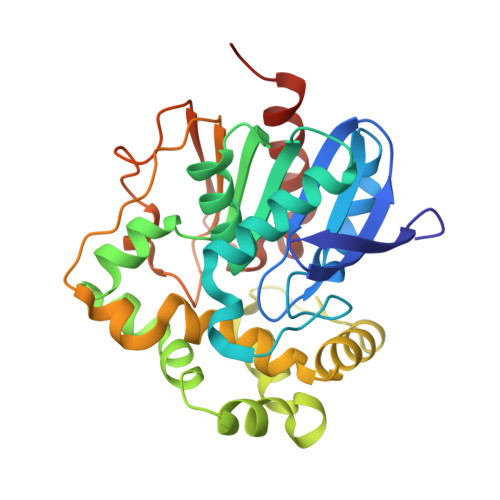
5NG7 | pdb_00005ng7

Novel epoxide hydrolases belonging to the alpha/beta hydrolases superfamily in metagenomes from hot environments


Experimental Data Snapshot

  • Method: X-RAY DIFFRACTION
  • Resolution: 1.39 Å
  • R-Value Free: 
    0.204 (Depositor), 0.203 (DCC) 
  • R-Value Work: 
    0.173 (Depositor) 
  • R-Value Observed: 
    0.174 (Depositor) 

Starting Model: experimental
View more details

wwPDB Validation 3D Report Full Report

Validation slider image for 5NG7

This is version 1.2 of the entry. See complete history. 

Literature

New Thermophilic alpha / beta Class Epoxide Hydrolases Found in Metagenomes From Hot Environments.

Ferrandi, E.E., Sayer, C., De Rose, S.A., Guazzelli, E., Marchesi, C., Saneei, V., Isupov, M.N., Littlechild, J.A., Monti, D.

(2018) Front Bioeng Biotechnol 6: 144-144

  • DOI: https://doi.org/10.3389/fbioe.2018.00144
  • Primary Citation Related Structures: 
    5NFQ, 5NG7

  • PubMed Abstract: 

    Two novel epoxide hydrolases (EHs), Sibe-EH and CH65-EH, were identified in the metagenomes of samples collected in hot springs in Russia and China, respectively. The two α/β hydrolase superfamily fold enzymes were cloned, over-expressed in Escherichia coli , purified and characterized. The new EHs were active toward a broad range of substrates, and in particular, Sibe-EH was excellent in the desymmetrization of cis -2,3-epoxybutane producing the (2 R ,3 R )-diol product with ee exceeding 99%. Interestingly these enzymes also hydrolyse (4 R )-limonene-1,2-epoxide with Sibe-EH being specific for the trans isomer. The Sibe-EH is a monomer in solution whereas the CH65-EH is a dimer. Both enzymes showed high melting temperatures with the CH65-EH being the highest at 85°C retaining 80% of its initial activity after 3 h thermal treatment at 70°C making it the most thermal tolerant wild type epoxide hydrolase described. The Sibe-EH and CH65-EH have been crystallized and their structures determined to high resolution, 1.6 and 1.4 Å, respectively. The CH65-EH enzyme forms a dimer via its cap domains with different relative orientation of the monomers compared to previously described EHs. The entrance to the active site cavity is located in a different position in CH65-EH and Sibe-EH in relation to other known bacterial and mammalian EHs.


  • Organizational Affiliation: 
    • Istituto di Chimica del Riconoscimento Molecolare, C.N.R., Milan, Italy.

Macromolecule Content 

  • Total Structure Weight: 70.38 kDa 
  • Atom Count: 6,046 
  • Modeled Residue Count: 582 
  • Deposited Residue Count: 598 
  • Unique protein chains: 1

Macromolecules

Find similar proteins by:|  3D Structure
Entity ID: 1
MoleculeChains  Sequence LengthOrganismDetailsImage
epoxide hydrolase
A, B
299metagenomeMutation(s): 0 
EC: 3.3.2.10
Entity Groups
Sequence Clusters30% Identity50% Identity70% Identity90% Identity95% Identity100% Identity
Sequence Annotations
Expand
Reference Sequence

Small Molecules

Ligands 4 Unique
IDChains Name / Formula / InChI Key2D Diagram3D Interactions
PEG

Query on PEG


Download:Ideal Coordinates CCD File
J [auth B]DI(HYDROXYETHYL)ETHER
C4 H10 O3
MTHSVFCYNBDYFN-UHFFFAOYSA-N
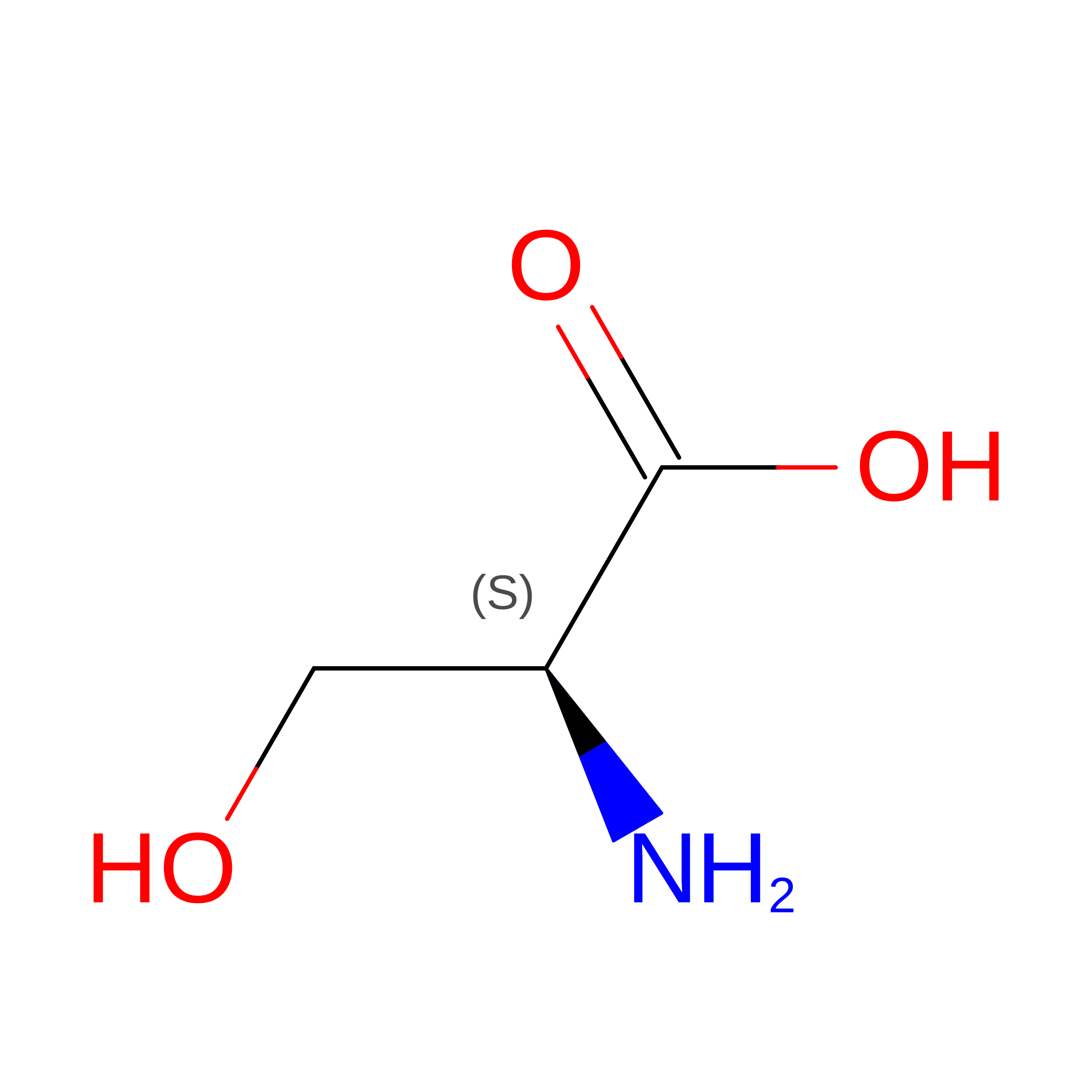
SER

Query on SER


Download:Ideal Coordinates CCD File
H [auth B]SERINE
C3 H7 N O3
MTCFGRXMJLQNBG-REOHCLBHSA-N
EDO

Query on EDO


Download:Ideal Coordinates CCD File
D [auth A],
E [auth A],
I [auth B]
1,2-ETHANEDIOL
C2 H6 O2
LYCAIKOWRPUZTN-UHFFFAOYSA-N
CL

Query on CL


Download:Ideal Coordinates CCD File
C [auth A],
F [auth B],
G [auth B]
CHLORIDE ION
Cl
VEXZGXHMUGYJMC-UHFFFAOYSA-M

Experimental Data & Validation

Experimental Data

  • Method: X-RAY DIFFRACTION
  • Resolution: 1.39 Å
  • R-Value Free:  0.204 (Depositor), 0.203 (DCC) 
  • R-Value Work:  0.173 (Depositor) 
  • R-Value Observed: 0.174 (Depositor) 
Space Group: C 1 2 1
Unit Cell:
Length ( Å )Angle ( ˚ )
a = 163.94α = 90
b = 46.22β = 106.94
c = 73.87γ = 90
Software Package:
Software NamePurpose
REFMACrefinement
XDSdata reduction
xia2data reduction
Aimlessdata scaling
MoRDaphasing

Structure Validation

View Full Validation Report


Entry History 

& Funding Information

Deposition Data


Funding OrganizationLocationGrant Number
European UnionUnited KingdomFP7/2007-2013 HOTZYME

Revision History  (Full details and data files)

  • Version 1.0: 2018-05-16
    Type: Initial release
  • Version 1.1: 2018-11-14
    Changes: Data collection, Database references
  • Version 1.2: 2024-01-17
    Changes: Data collection, Database references, Refinement description