5NEA

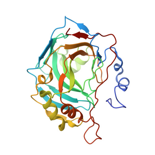
Crystal structure of human carbonic anhydrase II in complex with the inhibitor 4-(2-methyl-1,3-oxazol-5-yl)benzene-1-sulfonammide


Experimental Data Snapshot

  • Method: X-RAY DIFFRACTION
  • Resolution: 1.30 Å
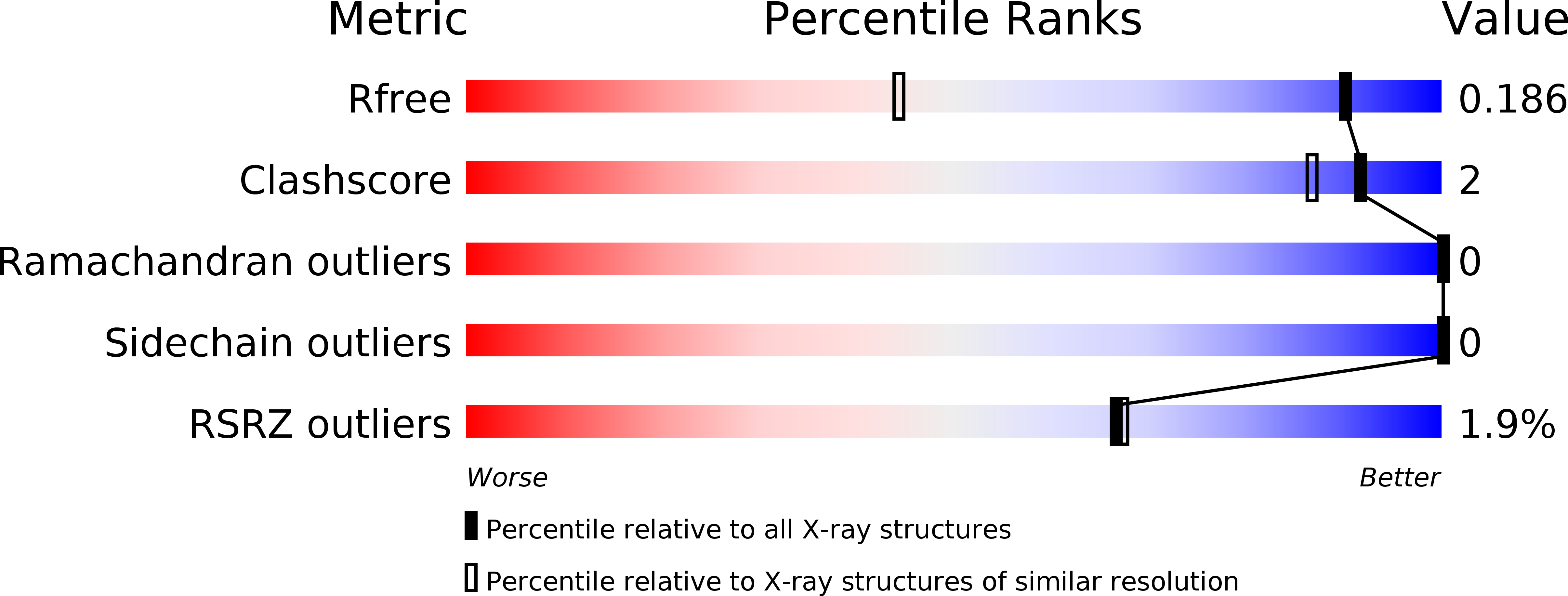
  • R-Value Free: 0.186 
  • R-Value Work: 0.166 
  • R-Value Observed: 0.167 

Starting Model: experimental
View more details

wwPDB Validation   3D Report Full Report


Ligand Structure Quality Assessment 


This is version 1.1 of the entry. See complete history


Literature

1,3-Oxazole-based selective picomolar inhibitors of cytosolic human carbonic anhydrase II alleviate ocular hypertension in rabbits: Potency is supported by X-ray crystallography of two leads.

Ferraroni, M.Lucarini, L.Masini, E.Korsakov, M.Scozzafava, A.Supuran, C.T.Krasavin, M.

(2017) Bioorg Med Chem 25: 4560-4565

  • DOI: https://doi.org/10.1016/j.bmc.2017.06.054
  • Primary Citation of Related Structures:  
    5NEA, 5NEE

  • PubMed Abstract: 

    Two lead 1,3-oxazole-based carbonic anhydrase inhibitors (CAIs) earlier identified as selective, picomolar inhibitors of hCA II (a cytosolic target for treatment of glaucoma) have been investigated further. Firstly, they were found to be conveniently synthesized on multigram scale, which enables further development. These compounds were found to be comparable in efficacy to dorzolamide eye drops when applied in the eye drop form as well. Finally, the reasons for unusually high potency of these compounds became understood from their high-resolution X-ray crystallography structures. These data significantly expand our understanding of heterocycle-based primary sulfonamides, many of which have recently emerged from our labs - particularly, from the corneal permeability standpoint.


  • Organizational Affiliation

    Chemistry Dept., Universita degli Studi di Firenze, Florence 50019, Italy.


Macromolecules
Find similar proteins by:  (by identity cutoff)  |  3D Structure
Entity ID: 1
MoleculeChains Sequence LengthOrganismDetailsImage
Carbonic anhydrase 2260Homo sapiensMutation(s): 0 
Gene Names: CA2
EC: 4.2.1.1 (PDB Primary Data), 4.2.1.69 (UniProt)
UniProt & NIH Common Fund Data Resources
Find proteins for P00918 (Homo sapiens)
Explore P00918 
Go to UniProtKB:  P00918
PHAROS:  P00918
GTEx:  ENSG00000104267 
Entity Groups  
Sequence Clusters30% Identity50% Identity70% Identity90% Identity95% Identity100% Identity
UniProt GroupP00918
Sequence Annotations
Expand
  • Reference Sequence
Small Molecules
Ligands 4 Unique
IDChains Name / Formula / InChI Key2D Diagram3D Interactions
8V8
Query on 8V8

Download Ideal Coordinates CCD File 
C [auth A],
D [auth A]
4-(2-methyl-1,3-oxazol-5-yl)benzenesulfonamide
C10 H10 N2 O3 S
FETRXICILYMBJH-UHFFFAOYSA-N
GOL
Query on GOL

Download Ideal Coordinates CCD File 
E [auth A]GLYCEROL
C3 H8 O3
PEDCQBHIVMGVHV-UHFFFAOYSA-N
ZN
Query on ZN

Download Ideal Coordinates CCD File 
B [auth A]ZINC ION
Zn
PTFCDOFLOPIGGS-UHFFFAOYSA-N
EDO
Query on EDO

Download Ideal Coordinates CCD File 
F [auth A]1,2-ETHANEDIOL
C2 H6 O2
LYCAIKOWRPUZTN-UHFFFAOYSA-N
Binding Affinity Annotations 
IDSourceBinding Affinity
8V8 BindingDB:  5NEA Ki: 0.05 (nM) from 1 assay(s)
Experimental Data & Validation

Experimental Data

  • Method: X-RAY DIFFRACTION
  • Resolution: 1.30 Å
  • R-Value Free: 0.186 
  • R-Value Work: 0.166 
  • R-Value Observed: 0.167 
  • Space Group: P 1 21 1
Unit Cell:
Length ( Å )Angle ( ˚ )
a = 42.38α = 90
b = 41.256β = 104.45
c = 72.071γ = 90
Software Package:
Software NamePurpose
XSCALEdata scaling
REFMACrefinement
PDB_EXTRACTdata extraction
XDSdata reduction
REFMACphasing

Structure Validation

View Full Validation Report


Ligand Structure Quality Assessment 


Entry History 

Deposition Data

Revision History  (Full details and data files)

  • Version 1.0: 2017-12-06
    Type: Initial release
  • Version 1.1: 2024-01-17
    Changes: Data collection, Database references, Refinement description