5NBD

PglK flippase in complex with inhibitory nanobody


Experimental Data Snapshot

  • Method: X-RAY DIFFRACTION
  • Resolution: 3.90 Å
  • R-Value Free: 0.335 
  • R-Value Work: 0.311 
  • R-Value Observed: 0.313 

Starting Models: experimental
View more details

wwPDB Validation   3D Report Full Report


Ligand Structure Quality Assessment 


This is version 1.1 of the entry. See complete history


Literature

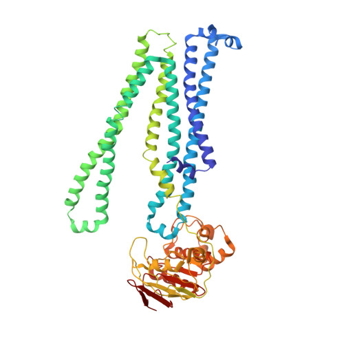
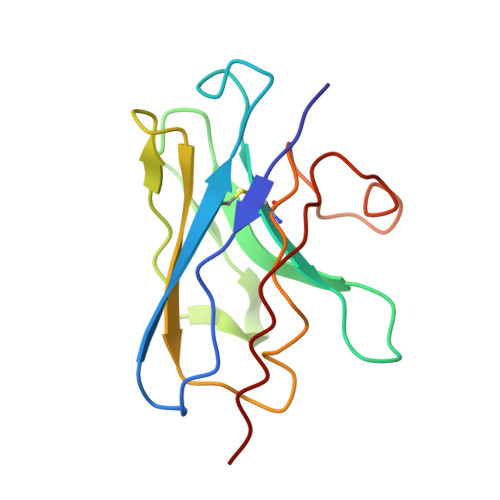
Structural basis of inhibition of lipid-linked oligosaccharide flippase PglK by a conformational nanobody.

Perez, C.Kohler, M.Janser, D.Pardon, E.Steyaert, J.Zenobi, R.Locher, K.P.

(2017) Sci Rep 7: 46641-46641

  • DOI: https://doi.org/10.1038/srep46641
  • Primary Citation of Related Structures:  
    5NBD

  • PubMed Abstract: 

    PglK is an ABC transporter that flips a lipid-linked oligosaccharide (LLO) that serves as a donor in protein N-glycosylation. Previous structures revealed two inward-facing conformations, both with very large separations of the nucleotide binding domains (NBDs), and a closed, ADP-bound state that featured an occluded cavity. To investigate additional states, we developed conformation-sensitive, single-domain camelid nanobodies (Nb) and studied their effect on PglK activity. Biochemical, structural, and mass spectrometric analyses revealed that one inhibitory Nb binds as a single copy to homodimeric PglK. The co-crystal structure of this Nb and ADP-bound PglK revealed a new, narrowly inward-open conformation. Rather than inducing asymmetry in the PglK homodimer, the binding of one Nb results in steric constraints that prevent a second Nb to access the symmetry-related site in PglK. The Nb performed its inhibitory role by a "sticky-doorstop" mechanism, where inhibition of ATP hydrolysis and LLO flipping activity occurs due to impaired closing of the NBD interface, which prevents PglK from converting to an outward-open conformation. This inhibitory mode suggests tight conformational coupling between the ATPase sites, which may apply to other ABC transporters.


  • Organizational Affiliation

    Department of Biology, Institute of Molecular Biology and Biophysics, ETH Zürich, CH-8093 Zürich, Switzerland.


Macromolecules
Find similar proteins by:  (by identity cutoff)  |  3D Structure
Entity ID: 1
MoleculeChains Sequence LengthOrganismDetailsImage
WlaB protein
A, B
564Campylobacter jejuniMutation(s): 1 
Gene Names: wlaB
Membrane Entity: Yes 
UniProt
Find proteins for Q0P9C4 (Campylobacter jejuni subsp. jejuni serotype O:2 (strain ATCC 700819 / NCTC 11168))
Explore Q0P9C4 
Go to UniProtKB:  Q0P9C4
Entity Groups  
Sequence Clusters30% Identity50% Identity70% Identity90% Identity95% Identity100% Identity
UniProt GroupQ0P9C4
Sequence Annotations
Expand
  • Reference Sequence
Find similar proteins by:  (by identity cutoff)  |  3D Structure
Entity ID: 2
MoleculeChains Sequence LengthOrganismDetailsImage
Nanobody123Lama glamaMutation(s): 0 
Entity Groups  
Sequence Clusters30% Identity50% Identity70% Identity90% Identity95% Identity100% Identity
Sequence Annotations
Expand
  • Reference Sequence
Small Molecules
Experimental Data & Validation

Experimental Data

  • Method: X-RAY DIFFRACTION
  • Resolution: 3.90 Å
  • R-Value Free: 0.335 
  • R-Value Work: 0.311 
  • R-Value Observed: 0.313 
  • Space Group: P 2 21 21
Unit Cell:
Length ( Å )Angle ( ˚ )
a = 84.34α = 90
b = 142.66β = 90
c = 199.48γ = 90
Software Package:
Software NamePurpose
XSCALEdata scaling
PHASERphasing
PHENIXrefinement
PDB_EXTRACTdata extraction
XDSdata reduction
PHASERphasing

Structure Validation

View Full Validation Report


Ligand Structure Quality Assessment 


Entry History & Funding Information

Deposition Data


Funding OrganizationLocationGrant Number
Swiss National Science FoundationSwitzerlandSNF 31003AB_131075 and 310030B_166672

Revision History  (Full details and data files)

  • Version 1.0: 2017-05-03
    Type: Initial release
  • Version 1.1: 2024-01-17
    Changes: Advisory, Data collection, Database references, Refinement description