5NA2 | pdb_00005na2

Crystal structure of the full-length Feline Immunodeficiency Virus capsid protein unveils original features


Experimental Data Snapshot

  • Method: X-RAY DIFFRACTION
  • Resolution: 1.67 Å
  • R-Value Free: 
    0.241 (Depositor), 0.243 (DCC) 
  • R-Value Work: 
    0.197 (Depositor), 0.200 (DCC) 
  • R-Value Observed: 
    0.199 (Depositor) 

Starting Models: experimental
View more details

wwPDB Validation 3D Report Full Report

Validation slider image for 5NA2

This is version 1.3 of the entry. See complete history

Literature

Crystal Structure of the Full-Length Feline Immunodeficiency Virus Capsid Protein Shows an N-Terminal beta-Hairpin in the Absence of N-Terminal Proline.

Folio, C.Sierra, N.Dujardin, M.Alvarez, G.Guillon, C.

(2017) Viruses 9

  • DOI: https://doi.org/10.3390/v9110335
  • Primary Citation Related Structures: 
    5NA2

  • PubMed Abstract: 

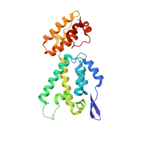
    Feline immunodeficiency virus (FIV) is a member of the Retroviridae family. It is the causative agent of an acquired immunodeficiency syndrome (AIDS) in cats and wild felines. Its capsid protein (CA) drives the assembly of the viral particle, which is a critical step in the viral replication cycle. Here, the first atomic structure of full-length FIV CA to 1.67 Å resolution is determined. The crystallized protein exhibits an original tetrameric assembly, composed of dimers which are stabilized by an intermolecular disulfide bridge induced by the crystallogenesis conditions. The FIV CA displays a standard α-helical CA topology with two domains, separated by a linker shorter than other retroviral CAs. The β-hairpin motif at its amino terminal end, which interacts with nucleotides in HIV-1, is unusually long in FIV CA. Interestingly, this functional β-motif is formed in this construct in the absence of the conserved N-terminal proline. The FIV CA exhibits a cis Arg-Pro bond in the CypA-binding loop, which is absent in known structures of lentiviral CAs. This structure represents the first tri-dimensional structure of a functional, full-length FIV CA.


  • Organizational Affiliation
    • Equipe Rétrovirus et Biochimie Structurale, Université de Lyon, CNRS, MMSB, UMR 5086 CNRS/Université de Lyon, IBCP, Lyon 69367 CEDEX 07, France. folio.christelle@gmail.com.

Macromolecule Content 

  • Total Structure Weight: 47.82 kDa 
  • Atom Count: 4,008 
  • Modeled Residue Count: 422 
  • Deposited Residue Count: 426 
  • Unique protein chains: 1

Macromolecules

Find similar proteins by:|  3D Structure
Entity ID: 1
MoleculeChains  Sequence LengthOrganismDetailsImage
Capsid protein (p24)
A, B
213Feline immunodeficiency virus (isolate Petaluma)Mutation(s): 1 
Gene Names: gag
UniProt
Find proteins for P16087 (Feline immunodeficiency virus (isolate Petaluma))
Explore P16087 
Go to UniProtKB:  P16087
Entity Groups
Sequence Clusters30% Identity50% Identity70% Identity90% Identity95% Identity100% Identity
UniProt GroupP16087
Sequence Annotations
Expand
Reference Sequence

Experimental Data & Validation

Experimental Data

  • Method: X-RAY DIFFRACTION
  • Resolution: 1.67 Å
  • R-Value Free:  0.241 (Depositor), 0.243 (DCC) 
  • R-Value Work:  0.197 (Depositor), 0.200 (DCC) 
  • R-Value Observed: 0.199 (Depositor) 
Space Group: C 1 2 1
Unit Cell:
Length ( Å )Angle ( ˚ )
a = 122.19α = 90
b = 74.58β = 128.68
c = 77γ = 90
Software Package:
Software NamePurpose
PHENIXrefinement
XDSdata reduction
XSCALEdata scaling
PHASERphasing

Structure Validation

View Full Validation Report


Entry History 

Deposition Data

Revision History  (Full details and data files)

  • Version 1.0: 2017-11-15
    Type: Initial release
  • Version 1.1: 2017-12-06
    Changes: Database references
  • Version 1.2: 2024-01-17
    Changes: Data collection, Database references, Refinement description
  • Version 1.3: 2024-11-20
    Changes: Structure summary