5N8O | pdb_00005n8o

Cryo EM structure of the conjugative relaxase TraI of the F/R1 plasmid system


Experimental Data Snapshot

  • Method: ELECTRON MICROSCOPY
  • Resolution: 3.90 Å
  • Aggregation State: PARTICLE 
  • Reconstruction Method: SINGLE PARTICLE 

wwPDB Validation 3D Report Full Report

Validation slider image for 5N8O

This is version 1.8 of the entry. See complete history

Literature

Cryo-EM Structure of a Relaxase Reveals the Molecular Basis of DNA Unwinding during Bacterial Conjugation

Ilangovan, A.Kay, C.W.M.Roier, S.El Mkami, H.Salvadori, E.Zechner, E.L.Zanetti, G.Waksman, G.

(2017) Cell 169: 708-721.e12

  • DOI: https://doi.org/10.1016/j.cell.2017.04.010
  • Primary Citation Related Structures: 
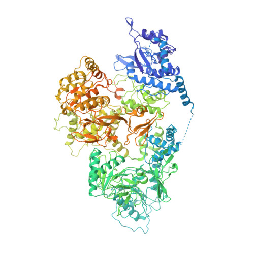
    5N8O

  • PubMed Abstract: 

    Relaxases play essential roles in conjugation, the main process by which bacteria exchange genetic material, notably antibiotic resistance genes. They are bifunctional enzymes containing a trans-esterase activity, which is responsible for nicking the DNA strand to be transferred and for covalent attachment to the resulting 5'-phosphate end, and a helicase activity, which is responsible for unwinding the DNA while it is being transported to a recipient cell. Here we show that these two activities are carried out by two conformers that can both load simultaneously on the origin of transfer DNA. We solve the structure of one of these conformers by cryo electron microscopy to near-atomic resolution, elucidating the molecular basis of helicase function by relaxases and revealing insights into the mechanistic events taking place in the cell prior to substrate transport during conjugation.


  • Organizational Affiliation
    • Institute of Structural and Molecular Biology, Birkbeck, Malet Street, London WC1E 7HX, UK.

Macromolecule Content 

  • Total Structure Weight: 198.64 kDa 
  • Atom Count: 11,362 
  • Modeled Residue Count: 1,450 
  • Deposited Residue Count: 1,778 
  • Unique protein chains: 1
  • Unique nucleic acid chains: 1

Macromolecules


Find similar proteins by:|  3D Structure
Entity ID: 1
MoleculeChains  Sequence LengthOrganismDetailsImage
DNA helicase I1,756Escherichia coliMutation(s): 0 
Gene Names: traI
EC: 5.6.2.3 (UniProt), 5.6.2 (UniProt)
UniProt
Find proteins for Q6TDU5 (Escherichia coli)
Explore Q6TDU5 
Go to UniProtKB:  Q6TDU5
Entity Groups
Sequence Clusters30% Identity50% Identity70% Identity90% Identity95% Identity100% Identity
UniProt GroupQ6TDU5
Sequence Annotations
Expand
Reference Sequence
Find similar nucleic acids by:  Sequence
Entity ID: 2
MoleculeChains LengthOrganismImage
DNA (5'-D(P*TP*TP*TP*TP*TP*TP*TP*TP*TP*TP*TP*TP*TP*TP*TP*TP*TP*T)-3')B [auth C]22Escherichia coli
Sequence Annotations
Expand
Reference Sequence

Experimental Data & Validation

Experimental Data

  • Method: ELECTRON MICROSCOPY
  • Resolution: 3.90 Å
  • Aggregation State: PARTICLE 
  • Reconstruction Method: SINGLE PARTICLE 
EM Software:
TaskSoftware PackageVersion
RECONSTRUCTIONRELION2.0
MODEL REFINEMENTPHENIX1.11.1

Structure Validation

View Full Validation Report


Entry History 

& Funding Information

Deposition Data


Funding OrganizationLocationGrant Number
Wellcome TrustUnited Kingdom--

Revision History  (Full details and data files)

  • Version 1.0: 2017-05-03
    Type: Initial release
  • Version 1.1: 2017-05-10
    Changes: Database references
  • Version 1.2: 2017-05-17
    Changes: Database references
  • Version 1.3: 2017-05-31
    Changes: Structure summary
  • Version 1.4: 2017-06-28
    Changes: Database references
  • Version 1.5: 2017-08-30
    Changes: Data collection, Experimental preparation
  • Version 1.6: 2019-12-11
    Changes: Other
  • Version 1.7: 2020-11-18
    Changes: Data collection
  • Version 1.8: 2024-05-15
    Changes: Data collection, Database references