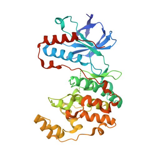
5N65

Crystal Structure of p38alpha in Complex with Lipid Pocket Ligand 9h


Experimental Data Snapshot

  • Method: X-RAY DIFFRACTION
  • Resolution: 2.00 Å
  • R-Value Free: 0.216 
  • R-Value Work: 0.192 
  • R-Value Observed: 0.193 

Starting Model: experimental
View more details

wwPDB Validation   3D Report Full Report


Ligand Structure Quality Assessment 


This is version 1.2 of the entry. See complete history


Literature

Structure-based design, synthesis and crystallization of 2-arylquinazolines as lipid pocket ligands of p38 alpha MAPK.

Buhrmann, M.Wiedemann, B.M.Muller, M.P.Hardick, J.Ecke, M.Rauh, D.

(2017) PLoS One 12: e0184627-e0184627

  • DOI: https://doi.org/10.1371/journal.pone.0184627
  • Primary Citation of Related Structures:  
    5N63, 5N64, 5N65, 5N66, 5N67, 5N68

  • PubMed Abstract: 

    In protein kinase research, identifying and addressing small molecule binding sites other than the highly conserved ATP-pocket are of intense interest because this line of investigation extends our understanding of kinase function beyond the catalytic phosphotransfer. Such alternative binding sites may be involved in altering the activation state through subtle conformational changes, control cellular enzyme localization, or in mediating and disrupting protein-protein interactions. Small organic molecules that target these less conserved regions might serve as tools for chemical biology research and to probe alternative strategies in targeting protein kinases in disease settings. Here, we present the structure-based design and synthesis of a focused library of 2-arylquinazoline derivatives to target the lipophilic C-terminal binding pocket in p38α MAPK, for which a clear biological function has yet to be identified. The interactions of the ligands with p38α MAPK was analyzed by SPR measurements and validated by protein X-ray crystallography.


  • Organizational Affiliation

    Faculty of Chemistry and Chemical Biology, TU Dortmund University, Dortmund, Germany.


Macromolecules
Find similar proteins by:  (by identity cutoff)  |  3D Structure
Entity ID: 1
MoleculeChains Sequence LengthOrganismDetailsImage
Mitogen-activated protein kinase 14360Homo sapiensMutation(s): 0 
Gene Names: MAPK14CSBPCSBP1CSBP2CSPB1MXI2SAPK2A
EC: 2.7.11.24
UniProt & NIH Common Fund Data Resources
Find proteins for Q16539 (Homo sapiens)
Explore Q16539 
Go to UniProtKB:  Q16539
PHAROS:  Q16539
GTEx:  ENSG00000112062 
Entity Groups  
Sequence Clusters30% Identity50% Identity70% Identity90% Identity95% Identity100% Identity
UniProt GroupQ16539
Sequence Annotations
Expand
  • Reference Sequence
Small Molecules
Experimental Data & Validation

Experimental Data

  • Method: X-RAY DIFFRACTION
  • Resolution: 2.00 Å
  • R-Value Free: 0.216 
  • R-Value Work: 0.192 
  • R-Value Observed: 0.193 
  • Space Group: P 21 21 21
Unit Cell:
Length ( Å )Angle ( ˚ )
a = 68.95α = 90
b = 69.74β = 90
c = 74.2γ = 90
Software Package:
Software NamePurpose
PHENIXrefinement
XDSdata reduction
XSCALEdata scaling
PHASERphasing

Structure Validation

View Full Validation Report


Ligand Structure Quality Assessment 


Entry History & Funding Information

Deposition Data


Funding OrganizationLocationGrant Number
German Federal Ministry for Education and Research (NGFNPlus and e:Med)GermanyBMBF 01GS08104, 01ZX1303C

Revision History  (Full details and data files)

  • Version 1.0: 2017-09-20
    Type: Initial release
  • Version 1.1: 2019-08-14
    Changes: Data collection
  • Version 1.2: 2024-01-17
    Changes: Data collection, Database references, Refinement description