5N4D | pdb_00005n4d

Prolyl oligopeptidase B from Galerina marginata bound to 25mer macrocyclization substrate - D661A mutant


Experimental Data Snapshot

  • Method: X-RAY DIFFRACTION
  • Resolution: 1.62 Å
  • R-Value Free: 
    0.234 (Depositor), 0.234 (DCC) 
  • R-Value Work: 
    0.198 (Depositor), 0.200 (DCC) 
  • R-Value Observed: 
    0.200 (Depositor) 

wwPDB Validation 3D Report Full Report

Validation slider image for 5N4D

This is version 2.3 of the entry. See complete history

Literature

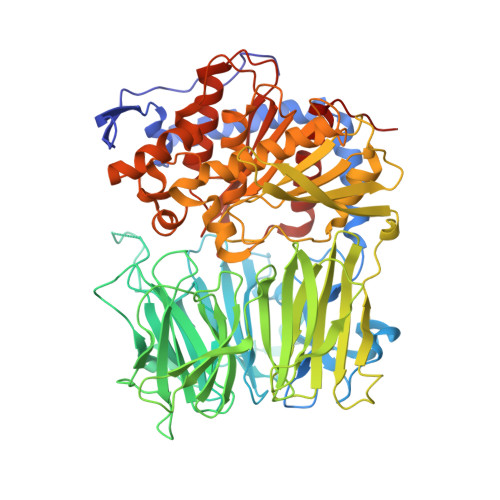
Characterization of a dual function macrocyclase enables design and use of efficient macrocyclization substrates.

Czekster, C.M.Ludewig, H.McMahon, S.A.Naismith, J.H.

(2017) Nat Commun 8: 1045-1045

  • DOI: https://doi.org/10.1038/s41467-017-00862-4
  • Primary Citation Related Structures: 
    5N4B, 5N4C, 5N4D, 5N4E, 5N4F

  • PubMed Abstract: 

    Peptide macrocycles are promising therapeutic molecules because they are protease resistant, structurally rigid, membrane permeable, and capable of modulating protein-protein interactions. Here, we report the characterization of the dual function macrocyclase-peptidase enzyme involved in the biosynthesis of the highly toxic amanitin toxin family of macrocycles. The enzyme first removes 10 residues from the N-terminus of a 35-residue substrate. Conformational trapping of the 25 amino-acid peptide forces the enzyme to release this intermediate rather than proceed to macrocyclization. The enzyme rebinds the 25 amino-acid peptide in a different conformation and catalyzes macrocyclization of the N-terminal eight residues. Structures of the enzyme bound to both substrates and biophysical analysis characterize the different binding modes rationalizing the mechanism. Using these insights simpler substrates with only five C-terminal residues were designed, allowing the enzyme to be more effectively exploited in biotechnology.


  • Organizational Affiliation
    • Biomedical Sciences Research Complex, The University of St Andrews, North Haugh, St Andrews, K16 9ST, UK.

Macromolecule Content 

  • Total Structure Weight: 169.44 kDa 
  • Atom Count: 13,347 
  • Modeled Residue Count: 1,478 
  • Deposited Residue Count: 1,510 
  • Unique protein chains: 2

Macromolecules

Find similar proteins by:|  3D Structure
Entity ID: 1
MoleculeChains  Sequence LengthOrganismDetailsImage
Prolyl oligopeptidase
A, B
730Galerina marginataMutation(s): 1 
Gene Names: POPB
EC: 3.4.21.26
UniProt
Find proteins for H2E7Q8 (Galerina marginata (strain CBS 339.88))
Explore H2E7Q8 
Go to UniProtKB:  H2E7Q8
Entity Groups
Sequence Clusters30% Identity50% Identity70% Identity90% Identity95% Identity100% Identity
UniProt GroupH2E7Q8
Sequence Annotations
Expand
Reference Sequence
Find similar proteins by:  Sequence   |   3D Structure  
Entity ID: 2
MoleculeChains  Sequence LengthOrganismDetailsImage
Alpha-amanitin proprotein
C, D
25Galerina marginataMutation(s): 0 
UniProt
Find proteins for A0A067SLB9 (Galerina marginata (strain CBS 339.88))
Explore A0A067SLB9 
Go to UniProtKB:  A0A067SLB9
Entity Groups
Sequence Clusters30% Identity50% Identity70% Identity90% Identity95% Identity100% Identity
UniProt GroupA0A067SLB9
Sequence Annotations
Expand
Reference Sequence

Experimental Data & Validation

Experimental Data

  • Method: X-RAY DIFFRACTION
  • Resolution: 1.62 Å
  • R-Value Free:  0.234 (Depositor), 0.234 (DCC) 
  • R-Value Work:  0.198 (Depositor), 0.200 (DCC) 
  • R-Value Observed: 0.200 (Depositor) 
Space Group: P 21 21 21
Unit Cell:
Length ( Å )Angle ( ˚ )
a = 99.04α = 90
b = 114.88β = 90
c = 141.31γ = 90
Software Package:
Software NamePurpose
PHENIXrefinement
xia2data reduction
Aimlessdata scaling
PHASERphasing

Structure Validation

View Full Validation Report


Entry History 

& Funding Information

Deposition Data


Funding OrganizationLocationGrant Number
European Research CouncilUnited Kingdom339367 NCB-TNT

Revision History  (Full details and data files)

  • Version 1.0: 2017-11-01
    Type: Initial release
  • Version 2.0: 2017-11-08
    Changes: Atomic model, Database references, Derived calculations
  • Version 2.1: 2018-01-24
    Changes: Source and taxonomy
  • Version 2.2: 2019-10-16
    Changes: Data collection
  • Version 2.3: 2024-05-08
    Changes: Data collection, Database references