5N38 | pdb_00005n38

S65DParkin and pUB complex


Experimental Data Snapshot

  • Method: X-RAY DIFFRACTION
  • Resolution: 2.60 Å
  • R-Value Free: 
    0.236 (Depositor), 0.249 (DCC) 
  • R-Value Work: 
    0.186 (Depositor), 0.197 (DCC) 
  • R-Value Observed: 
    0.189 (Depositor) 

Starting Models: experimental
View more details

wwPDB Validation 3D Report Full Report

Validation slider image for 5N38

This is version 1.6 of the entry. See complete history

Literature

Parkin-phosphoubiquitin complex reveals cryptic ubiquitin-binding site required for RBR ligase activity.

Kumar, A.Chaugule, V.K.Condos, T.E.C.Barber, K.R.Johnson, C.Toth, R.Sundaramoorthy, R.Knebel, A.Shaw, G.S.Walden, H.

(2017) Nat Struct Mol Biol 24: 475-483

  • DOI: https://doi.org/10.1038/nsmb.3400
  • Primary Citation Related Structures: 
    5N2W, 5N38

  • PubMed Abstract: 

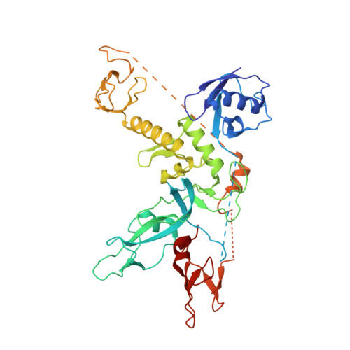
    RING-between-RING (RBR) E3 ligases are a class of ubiquitin ligases distinct from RING or HECT E3 ligases. An important RBR ligase is Parkin, mutations in which lead to early-onset hereditary Parkinsonism. Parkin and other RBR ligases share a catalytic RBR module but are usually autoinhibited and activated via distinct mechanisms. Recent insights into Parkin regulation predict large, unknown conformational changes during Parkin activation. However, current data on active RBR ligases reflect the absence of regulatory domains. Therefore, it remains unclear how individual RBR ligases are activated, and whether they share a common mechanism. We now report the crystal structure of a human Parkin-phosphoubiquitin complex, which shows that phosphoubiquitin binding induces movement in the 'in-between RING' (IBR) domain to reveal a cryptic ubiquitin-binding site. Mutation of this site negatively affects Parkin's activity. Furthermore, ubiquitin binding promotes cooperation between Parkin molecules, which suggests a role for interdomain association in the RBR ligase mechanism.


  • Organizational Affiliation
    • MRC Protein Phosphorylation and Ubiquitylation Unit, College of Life Sciences, University of Dundee, Dundee, UK.

Macromolecule Content 

  • Total Structure Weight: 54.93 kDa 
  • Atom Count: 3,633 
  • Modeled Residue Count: 455 
  • Deposited Residue Count: 481 
  • Unique protein chains: 2

Macromolecules

Find similar proteins by:|  3D Structure
Entity ID: 1
MoleculeChains  Sequence LengthOrganismDetailsImage
E3 ubiquitin-protein ligase parkin,E3 ubiquitin-protein ligase parkin405Homo sapiensMutation(s): 0 
Gene Names: PARK2PRKN
EC: 2.3.2 (PDB Primary Data), 2.3.2.31 (UniProt)
UniProt & NIH Common Fund Data Resources
Find proteins for O60260 (Homo sapiens)
Explore O60260 
Go to UniProtKB:  O60260
PHAROS:  O60260
GTEx:  ENSG00000185345 
Entity Groups
Sequence Clusters30% Identity50% Identity70% Identity90% Identity95% Identity100% Identity
UniProt GroupO60260
Sequence Annotations
Expand
Reference Sequence
Find similar proteins by:|  3D Structure
Entity ID: 2
MoleculeChains  Sequence LengthOrganismDetailsImage
Polyubiquitin-B76Homo sapiensMutation(s): 0 
Gene Names: UBB
UniProt & NIH Common Fund Data Resources
Find proteins for P0CG47 (Homo sapiens)
Explore P0CG47 
Go to UniProtKB:  P0CG47
PHAROS:  P0CG47
GTEx:  ENSG00000170315 
Entity Groups
Sequence Clusters30% Identity50% Identity70% Identity90% Identity95% Identity100% Identity
UniProt GroupP0CG47
Sequence Annotations
Expand
Reference Sequence

Small Molecules

Ligands 3 Unique
IDChains Name / Formula / InChI Key2D Diagram3D Interactions
PEG

Query on PEG


Download:Ideal Coordinates CCD File
C [auth A]DI(HYDROXYETHYL)ETHER
C4 H10 O3
MTHSVFCYNBDYFN-UHFFFAOYSA-N
ZN

Query on ZN


Download:Ideal Coordinates CCD File
E [auth A]
F [auth A]
G [auth A]
H [auth A]
I [auth A]
E [auth A],
F [auth A],
G [auth A],
H [auth A],
I [auth A],
J [auth A],
K [auth A],
L [auth A]
ZINC ION
Zn
PTFCDOFLOPIGGS-UHFFFAOYSA-N
CL

Query on CL


Download:Ideal Coordinates CCD File
D [auth A]CHLORIDE ION
Cl
VEXZGXHMUGYJMC-UHFFFAOYSA-M
Modified Residues  1 Unique
IDChains TypeFormula2D DiagramParent
SEP
Query on SEP
B
L-PEPTIDE LINKINGC3 H8 N O6 PSER

Experimental Data & Validation

Experimental Data

  • Method: X-RAY DIFFRACTION
  • Resolution: 2.60 Å
  • R-Value Free:  0.236 (Depositor), 0.249 (DCC) 
  • R-Value Work:  0.186 (Depositor), 0.197 (DCC) 
  • R-Value Observed: 0.189 (Depositor) 
Space Group: P 63 2 2
Unit Cell:
Length ( Å )Angle ( ˚ )
a = 146.54α = 90
b = 146.54β = 90
c = 88.44γ = 120
Software Package:
Software NamePurpose
BUSTERrefinement
MOSFLMdata reduction
Aimlessdata scaling
PHASERphasing

Structure Validation

View Full Validation Report


Entry History 

& Funding Information

Deposition Data


Funding OrganizationLocationGrant Number
Cancer Research UKUnited Kingdom17739
Medical Research Council (United Kingdom)United KingdomMC_UU_12016/12

Revision History  (Full details and data files)

  • Version 1.0: 2017-04-19
    Type: Initial release
  • Version 1.1: 2017-04-26
    Changes: Database references
  • Version 1.2: 2017-05-17
    Changes: Database references
  • Version 1.3: 2017-09-13
    Changes: Author supporting evidence
  • Version 1.4: 2018-02-07
    Changes: Database references
  • Version 1.5: 2024-01-17
    Changes: Data collection, Database references, Derived calculations, Refinement description
  • Version 1.6: 2024-11-13
    Changes: Derived calculations, Structure summary