5N10 | pdb_00005n10

Cucurbit[8]uril and 14-3-3 based binary bivalent supramolecular-protein assembly platform


Experimental Data Snapshot

  • Method: X-RAY DIFFRACTION
  • Resolution: 1.60 Å
  • R-Value Free: 
    0.194 (Depositor), 0.199 (DCC) 
  • R-Value Work: 
    0.170 (Depositor), 0.178 (DCC) 
  • R-Value Observed: 
    0.170 (Depositor) 

Starting Model: experimental
View more details

wwPDB Validation 3D Report Full Report

Validation slider image for 5N10

Ligand Structure Quality Assessment 


This is version 2.4 of the entry. See complete history

Literature

A Binary Bivalent Supramolecular Assembly Platform Based on Cucurbit[8]uril and Dimeric Adapter Protein 14-3-3.

de Vink, P.J.Briels, J.M.Schrader, T.Milroy, L.G.Brunsveld, L.Ottmann, C.

(2017) Angew Chem Int Ed Engl 56: 8998-9002

  • DOI: https://doi.org/10.1002/anie.201701807
  • Primary Citation Related Structures: 
    5N10

  • PubMed Abstract: 

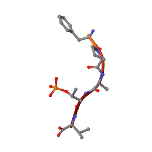
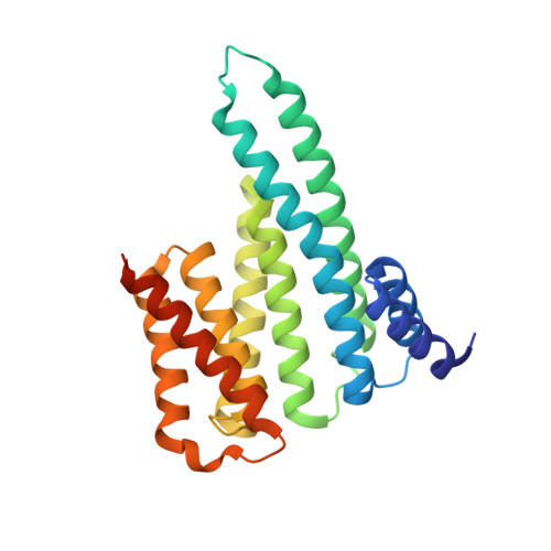
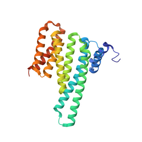
    Interactions between proteins frequently involve recognition sequences based on multivalent binding events. Dimeric 14-3-3 adapter proteins are a prominent example and typically bind partner proteins in a phosphorylation-dependent mono- or bivalent manner. Herein we describe the development of a cucurbit[8]uril (Q8)-based supramolecular system, which in conjunction with the 14-3-3 protein dimer acts as a binary and bivalent protein assembly platform. We fused the phenylalanine-glycine-glycine (FGG) tripeptide motif to the N-terminus of the 14-3-3-binding epitope of the estrogen receptor α (ERα) for selective binding to Q8. Q8-induced dimerization of the ERα epitope augmented its affinity towards 14-3-3 through a binary bivalent binding mode. The crystal structure of the Q8-induced ternary complex revealed molecular insight into the multiple supramolecular interactions between the protein, the peptide, and Q8.


  • Organizational Affiliation
    • Laboratory of Chemical Biology and Institute of Complex Molecular Systems, Department of Biomedical Engineering, Eindhoven University of Technology, Den Dolech 2, 5612 AZ, Eindhoven, The Netherlands.

Macromolecule Content 

  • Total Structure Weight: 62.52 kDa 
  • Atom Count: 4,894 
  • Modeled Residue Count: 490 
  • Deposited Residue Count: 540 
  • Unique protein chains: 3

Macromolecules

Find similar proteins by:  Sequence   |   3D Structure  
Entity ID: 1
MoleculeChains  Sequence LengthOrganismDetailsImage
Estrogen receptorA [auth F],
D [auth C],
E [auth D]
15Homo sapiensMutation(s): 0 
UniProt & NIH Common Fund Data Resources
Find proteins for P03372 (Homo sapiens)
Explore P03372 
Go to UniProtKB:  P03372
PHAROS:  P03372
GTEx:  ENSG00000091831 
Entity Groups
Sequence Clusters30% Identity50% Identity70% Identity90% Identity95% Identity100% Identity
UniProt GroupP03372
Sequence Annotations
Expand
Reference Sequence
Find similar proteins by:|  3D Structure
Entity ID: 2
MoleculeChains  Sequence LengthOrganismDetailsImage
14-3-3 protein beta/alphaB [auth A]246Homo sapiensMutation(s): 0 
Gene Names: YWHAB
UniProt & NIH Common Fund Data Resources
Find proteins for P31946 (Homo sapiens)
Explore P31946 
Go to UniProtKB:  P31946
PHAROS:  P31946
GTEx:  ENSG00000166913 
Entity Groups
Sequence Clusters30% Identity50% Identity70% Identity90% Identity95% Identity100% Identity
UniProt GroupP31946
Sequence Annotations
Expand
Reference Sequence
Find similar proteins by:|  3D Structure
Entity ID: 3
MoleculeChains  Sequence LengthOrganismDetailsImage
14-3-3 protein beta/alphaC [auth B]249Homo sapiensMutation(s): 0 
Gene Names: YWHAB
UniProt & NIH Common Fund Data Resources
Find proteins for P31946 (Homo sapiens)
Explore P31946 
Go to UniProtKB:  P31946
PHAROS:  P31946
GTEx:  ENSG00000166913 
Entity Groups
Sequence Clusters30% Identity50% Identity70% Identity90% Identity95% Identity100% Identity
UniProt GroupP31946
Sequence Annotations
Expand
Reference Sequence

Small Molecules

Modified Residues  1 Unique
IDChains TypeFormula2D DiagramParent
TPO
Query on TPO
A [auth F],
D [auth C],
E [auth D]
L-PEPTIDE LINKINGC4 H10 N O6 PTHR

Experimental Data & Validation

Experimental Data

  • Method: X-RAY DIFFRACTION
  • Resolution: 1.60 Å
  • R-Value Free:  0.194 (Depositor), 0.199 (DCC) 
  • R-Value Work:  0.170 (Depositor), 0.178 (DCC) 
  • R-Value Observed: 0.170 (Depositor) 
Space Group: C 1 2 1
Unit Cell:
Length ( Å )Angle ( ˚ )
a = 133.807α = 90
b = 57.571β = 98.64
c = 93.45γ = 90
Software Package:
Software NamePurpose
PHENIXrefinement
iMOSFLMdata reduction
Aimlessdata scaling
MOLREPphasing

Structure Validation

View Full Validation Report


Ligand Structure Quality Assessment 


Entry History 

& Funding Information

Deposition Data


Funding OrganizationLocationGrant Number
Netherlands Organization for Scientific ResearchNetherlands024.001.035
Netherlands Organization for Scientific ResearchNetherlands016.150.366
German Research FoundationGermanyCRC1093

Revision History  (Full details and data files)

  • Version 1.0: 2017-05-24
    Type: Initial release
  • Version 2.0: 2017-08-02
    Changes: Atomic model, Database references
  • Version 2.1: 2017-09-06
    Changes: Author supporting evidence
  • Version 2.2: 2019-10-16
    Changes: Data collection
  • Version 2.3: 2024-01-17
    Changes: Data collection, Database references, Refinement description
  • Version 2.4: 2024-11-06
    Changes: Structure summary