


X-ray structure of the H235Q mutant of GLIC in complex with bromoform
Fourati, Z., Delarue, M.To be published.
Experimental Data Snapshot
Entity ID: 1 | |||||
|---|---|---|---|---|---|
| Molecule | Chains | Sequence Length | Organism | Details | Image |
| Proton-gated ion channel | 327 | Gloeobacter violaceus PCC 7421 | Mutation(s): 1 Gene Names: glvI, glr4197 Membrane Entity: Yes | 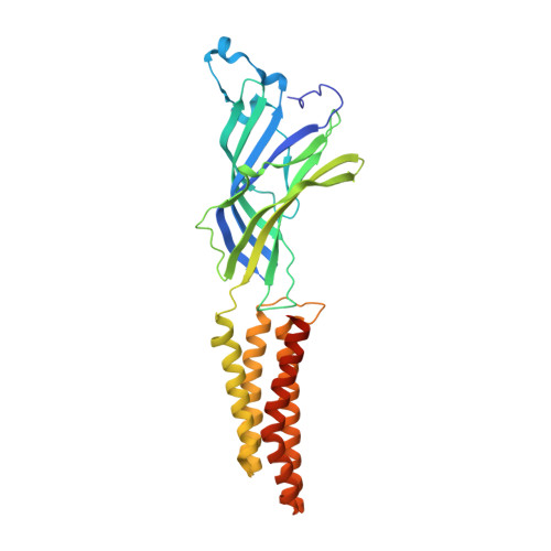 | |
UniProt | |||||
Entity Groups | |||||
| Sequence Clusters | 30% Identity50% Identity70% Identity90% Identity95% Identity100% Identity | ||||
| UniProt Group | Q7NDN8 | ||||
Sequence AnnotationsExpand | |||||
Reference Sequence | |||||
| Ligands 6 Unique | |||||
|---|---|---|---|---|---|
| ID | Chains | Name / Formula / InChI Key | 2D Diagram | 3D Interactions | |
| PLC Download:Ideal Coordinates CCD File | AB [auth E] BB [auth E] FA [auth C] G [auth A] GA [auth C] | DIUNDECYL PHOSPHATIDYL CHOLINE C32 H65 N O8 P IJFVSSZAOYLHEE-SSEXGKCCSA-O |  | ||
| LMT Download:Ideal Coordinates CCD File | BA [auth B] FB [auth E] LA [auth C] P [auth A] Q [auth A] | DODECYL-BETA-D-MALTOSIDE C24 H46 O11 NLEBIOOXCVAHBD-QKMCSOCLSA-N |  | ||
| MBR Download:Ideal Coordinates CCD File | CA [auth B] DA [auth B] GB [auth E] MA [auth C] NA [auth C] | TRIBROMOMETHANE C H Br3 DIKBFYAXUHHXCS-UHFFFAOYSA-N |  | ||
| ACT Download:Ideal Coordinates CCD File | AA [auth B] EA [auth C] EB [auth E] F [auth A] KA [auth C] | ACETATE ION C2 H3 O2 QTBSBXVTEAMEQO-UHFFFAOYSA-M |  | ||
| CL Download:Ideal Coordinates CCD File | CB [auth E] IA [auth C] J [auth A] K [auth A] L [auth A] | CHLORIDE ION Cl VEXZGXHMUGYJMC-UHFFFAOYSA-M |  | ||
| NA Download:Ideal Coordinates CCD File | DB [auth E] JA [auth C] M [auth A] N [auth A] TA [auth D] | SODIUM ION Na FKNQFGJONOIPTF-UHFFFAOYSA-N |  | ||
| Length ( Å ) | Angle ( ˚ ) |
|---|---|
| a = 181.87 | α = 90 |
| b = 132.764 | β = 102.43 |
| c = 160.292 | γ = 90 |
| Software Name | Purpose |
|---|---|
| BUSTER | refinement |
| XDS | data reduction |
| Aimless | data scaling |
| REFMAC | phasing |


