


Structural investigation of the interactions of ferrocene homobiotin derivative with avidin.
Strzelczyk, P., Plazuk, D., Bujacz, G.To be published.
Experimental Data Snapshot
Starting Model: experimental
View more details
Entity ID: 1 | |||||
|---|---|---|---|---|---|
| Molecule | Chains | Sequence Length | Organism | Details | Image |
| Avidin | 128 | Gallus gallus | Mutation(s): 0 | 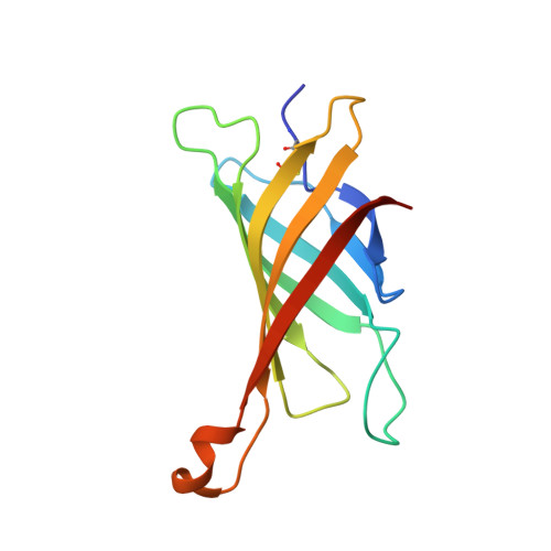 | |
UniProt | |||||
Entity Groups | |||||
| Sequence Clusters | 30% Identity50% Identity70% Identity90% Identity95% Identity100% Identity | ||||
| UniProt Group | P02701 | ||||
Glycosylation | |||||
| Glycosylation Sites: 1 | |||||
Sequence AnnotationsExpand | |||||
Reference Sequence | |||||
| Ligands 3 Unique | |||||
|---|---|---|---|---|---|
| ID | Chains | Name / Formula / InChI Key | 2D Diagram | 3D Interactions | |
| HBF Download:Ideal Coordinates CCD File | E [auth A], K [auth B], S [auth C], Z [auth D] | ferrocene homobiotin derivative C21 H17 Fe N2 O S YYWARPGDAKPMLM-VZXRKJOKSA-N |  | ||
| NAG Download:Ideal Coordinates CCD File | AA [auth D], F [auth A], L [auth B], T [auth C] | 2-acetamido-2-deoxy-beta-D-glucopyranose C8 H15 N O6 OVRNDRQMDRJTHS-FMDGEEDCSA-N |  | ||
| IOD Download:Ideal Coordinates CCD File | BA [auth D] CA [auth D] DA [auth D] EA [auth D] FA [auth D] | IODIDE ION I XMBWDFGMSWQBCA-UHFFFAOYSA-M |  | ||
| Length ( Å ) | Angle ( ˚ ) |
|---|---|
| a = 43.933 | α = 90 |
| b = 82.081 | β = 106.29 |
| c = 74.601 | γ = 90 |
| Software Name | Purpose |
|---|---|
| REFMAC | refinement |
| XDS | data reduction |
| MOLREP | phasing |


