5MYB

Homodimerization of Tie2 Fibronectin-like domains 2 and 3 in space group P21


Experimental Data Snapshot

  • Method: X-RAY DIFFRACTION
  • Resolution: 2.60 Å
  • R-Value Free: 0.278 
  • R-Value Work: 0.222 
  • R-Value Observed: 0.225 

Starting Model: experimental
View more details

wwPDB Validation   3D Report Full Report


Ligand Structure Quality Assessment 


This is version 2.1 of the entry. See complete history


Literature

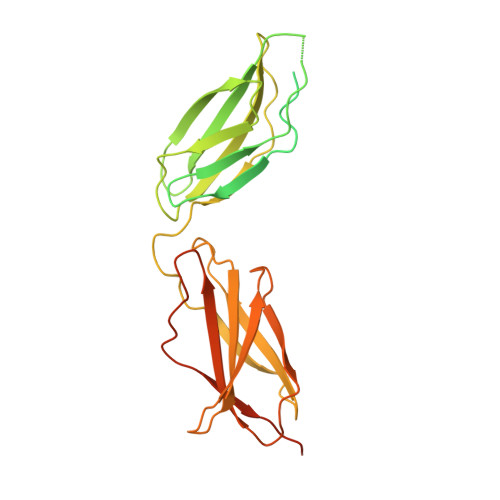
Structural basis of Tie2 activation and Tie2/Tie1 heterodimerization.

Leppanen, V.M.Saharinen, P.Alitalo, K.

(2017) Proc Natl Acad Sci U S A 114: 4376-4381

  • DOI: https://doi.org/10.1073/pnas.1616166114
  • Primary Citation of Related Structures:  
    5MYA, 5MYB, 5N06

  • PubMed Abstract: 

    The endothelial cell (EC)-specific receptor tyrosine kinases Tie1 and Tie2 are necessary for the remodeling and maturation of blood and lymphatic vessels. Angiopoietin-1 (Ang1) growth factor is a Tie2 agonist, whereas Ang2 functions as a context-dependent agonist/antagonist. The orphan receptor Tie1 modulates Tie2 activation, which is induced by association of angiopoietins with Tie2 in cis and across EC-EC junctions in trans Except for the binding of the C-terminal angiopoietin domains to the Tie2 ligand-binding domain, the mechanisms for Tie2 activation are poorly understood. We report here the structural basis of Ang1-induced Tie2 dimerization in cis and provide mechanistic insights on Ang2 antagonism, Tie1/Tie2 heterodimerization, and Tie2 clustering. We find that Ang1-induced Tie2 dimerization and activation occurs via the formation of an intermolecular β-sheet between the membrane-proximal (third) Fibronectin type III domains (Fn3) of Tie2. The structures of Tie2 and Tie1 Fn3 domains are similar and compatible with Tie2/Tie1 heterodimerization by the same mechanism. Mutagenesis of the key interaction residues of Tie2 and Tie1 Fn3 domains decreased Ang1-induced Tie2 phosphorylation and increased the basal phosphorylation of Tie1, respectively. Furthermore, the Tie2 structures revealed additional interactions between the Fn 2 (Fn2) domains that coincide with a mutation of Tie2 in primary congenital glaucoma that leads to defective Tie2 clustering and junctional localization. Mutagenesis of the Fn2-Fn2 interface increased the basal phosphorylation of Tie2, suggesting that the Fn2 interactions are essential in preformed Tie2 oligomerization. The interactions of the membrane-proximal domains could provide new targets for modulation of Tie receptor activity.


  • Organizational Affiliation

    Wihuri Research Institute, Biomedicum Helsinki, Haartmaninkatu 8, 00290 Helsinki, Finland; kari.alitalo@helsinki.fi veli-matti.leppanen@helsinki.fi.


Macromolecules
Find similar proteins by:  (by identity cutoff)  |  3D Structure
Entity ID: 1
MoleculeChains Sequence LengthOrganismDetailsImage
Angiopoietin-1 receptor
A, B
333Homo sapiensMutation(s): 0 
Gene Names: TEKTIE2VMCMVMCM1
EC: 2.7.10.1
UniProt & NIH Common Fund Data Resources
Find proteins for Q02763 (Homo sapiens)
Explore Q02763 
Go to UniProtKB:  Q02763
GTEx:  ENSG00000120156 
Entity Groups  
Sequence Clusters30% Identity50% Identity70% Identity90% Identity95% Identity100% Identity
UniProt GroupQ02763
Glycosylation
Glycosylation Sites: 3Go to GlyGen: Q02763-1
Sequence Annotations
Expand
  • Reference Sequence
Oligosaccharides

Help

Entity ID: 2
MoleculeChains Length2D Diagram Glycosylation3D Interactions
beta-D-mannopyranose-(1-3)-2-acetamido-2-deoxy-beta-D-glucopyranose-(1-4)-2-acetamido-2-deoxy-beta-D-glucopyranose
C
3N-Glycosylation
Glycosylation Resources
GlyTouCan:  G44266EX
GlyCosmos:  G44266EX
GlyGen:  G44266EX
Entity ID: 3
MoleculeChains Length2D Diagram Glycosylation3D Interactions
2-acetamido-2-deoxy-beta-D-glucopyranose-(1-4)-2-acetamido-2-deoxy-beta-D-glucopyranose
D
2N-Glycosylation
Glycosylation Resources
GlyTouCan:  G42666HT
GlyCosmos:  G42666HT
GlyGen:  G42666HT
Experimental Data & Validation

Experimental Data

  • Method: X-RAY DIFFRACTION
  • Resolution: 2.60 Å
  • R-Value Free: 0.278 
  • R-Value Work: 0.222 
  • R-Value Observed: 0.225 
  • Space Group: P 1 21 1
Unit Cell:
Length ( Å )Angle ( ˚ )
a = 78.46α = 90
b = 48.49β = 111.09
c = 80.67γ = 90
Software Package:
Software NamePurpose
PHENIXrefinement
XDSdata reduction
XSCALEdata scaling
autoSHARPphasing

Structure Validation

View Full Validation Report


Ligand Structure Quality Assessment 


Entry History 

Deposition Data

Revision History  (Full details and data files)

  • Version 1.0: 2017-04-26
    Type: Initial release
  • Version 1.1: 2017-05-03
    Changes: Database references
  • Version 2.0: 2020-07-29
    Type: Remediation
    Reason: Carbohydrate remediation
    Changes: Advisory, Atomic model, Data collection, Derived calculations, Structure summary
  • Version 2.1: 2024-05-01
    Changes: Data collection, Database references, Derived calculations, Refinement description, Structure summary