5MX9 | pdb_00005mx9

High resolution crystal structure of the MCR-2 catalytic domain


Experimental Data Snapshot

  • Method: X-RAY DIFFRACTION
  • Resolution: 1.12 Å
  • R-Value Free: 
    0.145 (Depositor), 0.146 (DCC) 
  • R-Value Work: 
    0.133 (Depositor), 0.135 (DCC) 
  • R-Value Observed: 
    0.134 (Depositor) 

Starting Model: experimental
View more details

wwPDB Validation 3D Report Full Report

Validation slider image for 5MX9

This is version 1.3 of the entry. See complete history

Literature

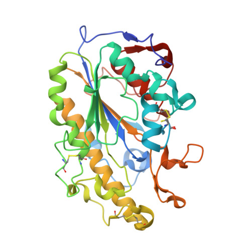
1.12 angstrom resolution crystal structure of the catalytic domain of the plasmid-mediated colistin resistance determinant MCR-2.

Coates, K.Walsh, T.R.Spencer, J.Hinchliffe, P.

(2017) Acta Crystallogr F Struct Biol Commun 73: 443-449

  • DOI: https://doi.org/10.1107/S2053230X17009669
  • Primary Citation Related Structures: 
    5MX9

  • PubMed Abstract: 

    MCR-2 confers resistance to colistin, a `last-line' antibiotic against extensively resistant Gram-negative pathogens. It is a plasmid-encoded phosphoethanolamine transferase that is closely related to MCR-1. To understand the diversity in the MCR family, the 1.12 Å resolution crystal structure of the catalytic domain of MCR-2 was determined. Variable amino acids are located distant from both the di-zinc active site and the membrane-proximal face. The exceptionally high resolution will provide an accurate starting model for further mechanistic studies.


  • Organizational Affiliation
    • School of Cellular and Molecular Medicine, University of Bristol, Bristol BS8 1TD, England.

Macromolecule Content 

  • Total Structure Weight: 36.49 kDa 
  • Atom Count: 3,004 
  • Modeled Residue Count: 324 
  • Deposited Residue Count: 324 
  • Unique protein chains: 1

Macromolecules

Find similar proteins by:|  3D Structure
Entity ID: 1
MoleculeChains  Sequence LengthOrganismDetailsImage
Phosphatidylethanolamine transferase Mcr-2324Escherichia coliMutation(s): 0 
Gene Names: mcr-2
EC: 2.7.8.43
UniProt
Find proteins for A0A1C3NEV1 (Escherichia coli)
Explore A0A1C3NEV1 
Go to UniProtKB:  A0A1C3NEV1
Entity Groups
Sequence Clusters30% Identity50% Identity70% Identity90% Identity95% Identity100% Identity
UniProt GroupA0A1C3NEV1
Sequence Annotations
Expand
Reference Sequence

Experimental Data & Validation

Experimental Data

  • Method: X-RAY DIFFRACTION
  • Resolution: 1.12 Å
  • R-Value Free:  0.145 (Depositor), 0.146 (DCC) 
  • R-Value Work:  0.133 (Depositor), 0.135 (DCC) 
  • R-Value Observed: 0.134 (Depositor) 
Space Group: P 21 21 21
Unit Cell:
Length ( Å )Angle ( ˚ )
a = 44.818α = 90
b = 53.31β = 90
c = 117.509γ = 90
Software Package:
Software NamePurpose
PHENIXrefinement
DIALSdata reduction
Aimlessdata scaling
PHASERphasing

Structure Validation

View Full Validation Report


Entry History 

& Funding Information

Deposition Data


Funding OrganizationLocationGrant Number
Medical Research Council (United Kingdom)United KingdomMR/P007295/1

Revision History  (Full details and data files)

  • Version 1.0: 2017-08-09
    Type: Initial release
  • Version 1.1: 2017-08-23
    Changes: Database references
  • Version 1.2: 2024-01-17
    Changes: Data collection, Database references, Derived calculations, Refinement description
  • Version 1.3: 2024-10-23
    Changes: Structure summary