5MX7

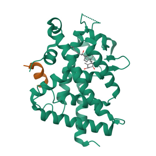
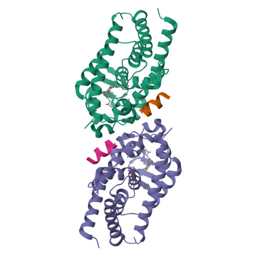
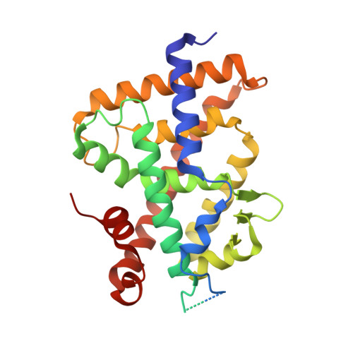
1a,20S-dihydroxyvitamin D3 VDR complex


Experimental Data Snapshot

  • Method: X-RAY DIFFRACTION
  • Resolution: 1.98 Å
  • R-Value Free: 0.225 
  • R-Value Work: 0.200 
  • R-Value Observed: 0.201 

Starting Model: experimental
View more details

wwPDB Validation   3D Report Full Report


Ligand Structure Quality Assessment 


This is version 1.2 of the entry. See complete history


Literature

1 alpha,20S-Dihydroxyvitamin D3 Interacts with Vitamin D Receptor: Crystal Structure and Route of Chemical Synthesis.

Lin, Z.Chen, H.Belorusova, A.Y.Bollinger, J.C.Tang, E.K.Y.Janjetovic, Z.Kim, T.K.Wu, Z.Miller, D.D.Slominski, A.T.Postlethwaite, A.E.Tuckey, R.C.Rochel, N.Li, W.

(2017) Sci Rep 7: 10193-10193

  • DOI: https://doi.org/10.1038/s41598-017-10917-7
  • Primary Citation of Related Structures:  
    5MX7

  • PubMed Abstract: 

    1α,20S-Dihydroxyvitamin D3 [1,20S(OH) 2 D 3 ], a natural and bioactive vitamin D3 metabolite, was chemically synthesized for the first time. X-ray crystallography analysis of intermediate 15 confirmed its 1α-OH configuration. 1,20S(OH) 2 D 3 interacts with the vitamin D receptor (VDR), with similar potency to its native ligand, 1α,25-dihydroxyvitamin D 3 [1,25(OH) 2 D 3 ] as illustrated by its ability to stimulate translocation of the VDR to the nucleus, stimulate VDRE-reporter activity, regulate VDR downstream genes (VDR, CYP24A1, TRPV6 and CYP27B1), and inhibit the production of inflammatory markers (IFNγ and IL1β). However, their co-crystal structures revealed differential molecular interactions of the 20S-OH moiety and the 25-OH moiety to the VDR, which may explain some differences in their biological activities. Furthermore, this study provides a synthetic route for the synthesis of 1,20S(OH) 2 D 3 using the intermediate 1α,3β-diacetoxypregn-5-en-20-one (3), and provides a molecular and biological basis for the development of 1,20S(OH) 2 D 3 and its analogs as potential therapeutic agents.


  • Organizational Affiliation

    Department of Pharmaceutical Sciences, University of Tennessee Health Science Center, 881 Madison Avenue, Room 561, Memphis, TN, 38163, United States.


Macromolecules
Find similar proteins by:  (by identity cutoff)  |  3D Structure
Entity ID: 1
MoleculeChains Sequence LengthOrganismDetailsImage
Vitamin D3 receptor AA [auth A1]300Danio rerioMutation(s): 0 
Gene Names: vdranr1i1avdr
UniProt
Find proteins for Q9PTN2 (Danio rerio)
Explore Q9PTN2 
Go to UniProtKB:  Q9PTN2
Entity Groups  
Sequence Clusters30% Identity50% Identity70% Identity90% Identity95% Identity100% Identity
UniProt GroupQ9PTN2
Sequence Annotations
Expand
  • Reference Sequence

Find similar proteins by:  Sequence   |   3D Structure  

Entity ID: 2
MoleculeChains Sequence LengthOrganismDetailsImage
Nuclear receptor coactivator 1B [auth B1]15Homo sapiensMutation(s): 0 
EC: 2.3.1.48
UniProt & NIH Common Fund Data Resources
Find proteins for Q15788 (Homo sapiens)
Explore Q15788 
Go to UniProtKB:  Q15788
PHAROS:  Q15788
GTEx:  ENSG00000084676 
Entity Groups  
Sequence Clusters30% Identity50% Identity70% Identity90% Identity95% Identity100% Identity
UniProt GroupQ15788
Sequence Annotations
Expand
  • Reference Sequence
Small Molecules
Ligands 1 Unique
IDChains Name / Formula / InChI Key2D Diagram3D Interactions
D3V
Query on D3V

Download Ideal Coordinates CCD File 
C [auth A1] 1a,20S-dihydroxyvitamin D3
C27 H44 O3
RLDKSDBKVGPMKA-YAUUYYILSA-N
Experimental Data & Validation

Experimental Data

  • Method: X-RAY DIFFRACTION
  • Resolution: 1.98 Å
  • R-Value Free: 0.225 
  • R-Value Work: 0.200 
  • R-Value Observed: 0.201 
  • Space Group: P 65 2 2
Unit Cell:
Length ( Å )Angle ( ˚ )
a = 65.936α = 90
b = 65.936β = 90
c = 262.97γ = 120
Software Package:
Software NamePurpose
PHENIXrefinement
HKL-2000data reduction
HKL-2000data scaling
PHENIXphasing

Structure Validation

View Full Validation Report


Ligand Structure Quality Assessment 


Entry History & Funding Information

Deposition Data


Funding OrganizationLocationGrant Number
French National Research AgencyFranceANR-13-BSV8-0024-01

Revision History  (Full details and data files)

  • Version 1.0: 2017-11-29
    Type: Initial release
  • Version 1.1: 2021-02-24
    Changes: Derived calculations
  • Version 1.2: 2024-01-17
    Changes: Data collection, Database references, Refinement description