5MX6 | pdb_00005mx6

Crystal structure of H. pylori purine nucleoside phosphorylase from clinical isolate HpPNP-2


Experimental Data Snapshot

  • Method: X-RAY DIFFRACTION
  • Resolution: 2.41 Å
  • R-Value Free: 
    0.225 (Depositor), 0.225 (DCC) 
  • R-Value Work: 
    0.175 (Depositor), 0.175 (DCC) 
  • R-Value Observed: 
    0.177 (Depositor) 

Starting Model: experimental
View more details

wwPDB Validation 3D Report Full Report

Validation slider image for 5MX6

This is version 1.3 of the entry. See complete history

Literature

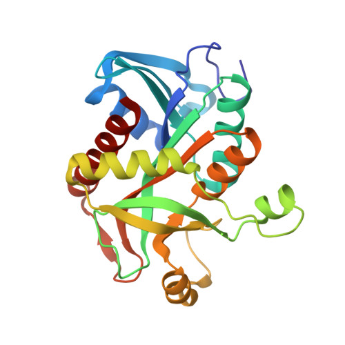
Structural characterization of purine nucleoside phosphorylase from human pathogen Helicobacter pylori.

Stefanic, Z.Mikleusevic, G.Luic, M.Bzowska, A.Lescic Asler, I.

(2017) Int J Biol Macromol 101: 518-526

  • DOI: https://doi.org/10.1016/j.ijbiomac.2017.03.101
  • Primary Citation Related Structures: 
    5MX4, 5MX6, 5MX8

  • PubMed Abstract: 

    Microaerophilic bacterium Helicobacer pylori is a well known human pathogen involved in the development of many diseases. Due to the evergrowing infection rate and increase of H. pylori antibiotic resistence, it is of utmost importance to find a new way to attack and eradicate H. pylori. The purine metabolism in H. pylori is solely dependant on the salvage pathway and one of the key enzymes in this pathway is purine nucleoside phosphorylase (PNP). In this timely context, we report here the basic biochemical and structural characterization of recombinant PNP from the H. pylori clinical isolate expressed in Escherichia coli. Structure of H. pylori PNP is typical for high molecular mass PNPs. However, its activity towards adenosine is very low, thus resembling more that of low molecular mass PNPs. Understanding the molecular mechanism of this key enzyme may lead to the development of new drug strategies and help in the eradication of H. pylori.


  • Organizational Affiliation
    • Division of Physical Chemistry, Ruđer Bošković Institute, POB 180, Bijenička cesta 54, HR-10002 Zagreb, Croatia.

Macromolecule Content 

  • Total Structure Weight: 157.33 kDa 
  • Atom Count: 11,499 
  • Modeled Residue Count: 1,398 
  • Deposited Residue Count: 1,398 
  • Unique protein chains: 1

Macromolecules

Find similar proteins by:|  3D Structure
Entity ID: 1
MoleculeChains  Sequence LengthOrganismDetailsImage
Purine nucleoside phosphorylase DeoD-type
A, B, C, D, E
A, B, C, D, E, F
233Helicobacter pyloriMutation(s): 0 
EC: 2.4.2.1
UniProt
Find proteins for P56463 (Helicobacter pylori (strain ATCC 700392 / 26695))
Explore P56463 
Go to UniProtKB:  P56463
Entity Groups
Sequence Clusters30% Identity50% Identity70% Identity90% Identity95% Identity100% Identity
UniProt GroupP56463
Sequence Annotations
Expand
Reference Sequence

Small Molecules

Ligands 5 Unique
IDChains Name / Formula / InChI Key2D Diagram3D Interactions
HPA

Query on HPA


Download:Ideal Coordinates CCD File
AA [auth F]
H [auth A]
M [auth B]
P [auth C]
T [auth D]
AA [auth F],
H [auth A],
M [auth B],
P [auth C],
T [auth D],
X [auth E]
HYPOXANTHINE
C5 H4 N4 O
FDGQSTZJBFJUBT-UHFFFAOYSA-N
PEG

Query on PEG


Download:Ideal Coordinates CCD File
K [auth A],
N [auth B],
R [auth C],
V [auth D],
Y [auth E]
DI(HYDROXYETHYL)ETHER
C4 H10 O3
MTHSVFCYNBDYFN-UHFFFAOYSA-N
SO4

Query on SO4


Download:Ideal Coordinates CCD File
BA [auth F]
G [auth A]
I [auth A]
L [auth B]
Q [auth C]
BA [auth F],
G [auth A],
I [auth A],
L [auth B],
Q [auth C],
S [auth D],
U [auth D],
W [auth E],
Z [auth F]
SULFATE ION
O4 S
QAOWNCQODCNURD-UHFFFAOYSA-L
PO4

Query on PO4


Download:Ideal Coordinates CCD File
O [auth C]PHOSPHATE ION
O4 P
NBIIXXVUZAFLBC-UHFFFAOYSA-K
ACT

Query on ACT


Download:Ideal Coordinates CCD File
CA [auth F],
J [auth A]
ACETATE ION
C2 H3 O2
QTBSBXVTEAMEQO-UHFFFAOYSA-M

Experimental Data & Validation

Experimental Data

  • Method: X-RAY DIFFRACTION
  • Resolution: 2.41 Å
  • R-Value Free:  0.225 (Depositor), 0.225 (DCC) 
  • R-Value Work:  0.175 (Depositor), 0.175 (DCC) 
  • R-Value Observed: 0.177 (Depositor) 
Space Group: P 21 21 21
Unit Cell:
Length ( Å )Angle ( ˚ )
a = 104.26α = 90
b = 119.86β = 90
c = 139.22γ = 90
Software Package:
Software NamePurpose
PHENIXrefinement
SCALAdata scaling
PDB_EXTRACTdata extraction
MOSFLMdata reduction
PHASERphasing

Structure Validation

View Full Validation Report


Entry History 

Deposition Data

  • Released Date: 2017-04-05 
  • Deposition Author(s): Stefanic, Z.

Revision History  (Full details and data files)

  • Version 1.0: 2017-04-05
    Type: Initial release
  • Version 1.1: 2017-04-12
    Changes: Database references
  • Version 1.2: 2024-01-17
    Changes: Data collection, Database references, Refinement description
  • Version 1.3: 2025-10-01
    Changes: Advisory, Derived calculations, Structure summary