5MX1 | pdb_00005mx1

Crystal structure of human chondroadherin


Experimental Data Snapshot

  • Method: X-RAY DIFFRACTION
  • Resolution: 2.17 Å
  • R-Value Free: 
    0.239 (Depositor), 0.240 (DCC) 
  • R-Value Work: 
    0.188 (Depositor), 0.194 (DCC) 
  • R-Value Observed: 
    0.191 (Depositor) 

Starting Model: experimental
View more details

wwPDB Validation 3D Report Full Report

Validation slider image for 5MX1

This is version 1.3 of the entry. See complete history

Literature

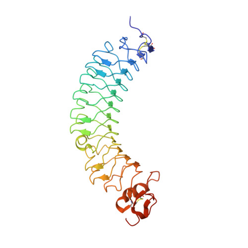
Structural and functional analysis of two small leucine-rich repeat proteoglycans, fibromodulin and chondroadherin.

Paracuellos, P.Kalamajski, S.Bonna, A.Bihan, D.Farndale, R.W.Hohenester, E.

(2017) Matrix Biol 63: 106-116

  • DOI: https://doi.org/10.1016/j.matbio.2017.02.002
  • Primary Citation Related Structures: 
    5MX0, 5MX1

  • PubMed Abstract: 

    The small leucine-rich proteoglycans (SLRPs) are important regulators of extracellular matrix assembly and cell signalling. We have determined crystal structures at ~2.2Å resolution of human fibromodulin and chondroadherin, two collagen-binding SLRPs. Their overall fold is similar to that of the prototypical SLRP, decorin, but unlike decorin neither fibromodulin nor chondroadherin forms a stable dimer. A previously identified binding site for integrin α2β1 maps to an α-helix in the C-terminal cap region of chondroadherin. Interrogation of the Collagen Toolkits revealed a unique binding site for chondroadherin in collagen II, and no binding to collagen III. A triple-helical peptide containing the sequence GAOGPSGFQGLOGPOGPO (O is hydroxyproline) forms a stable complex with chondroadherin in solution. In fibrillar collagen I and II, this sequence is aligned with the collagen cross-linking site KGHR, suggesting a role for chondroadherin in cross-linking.


  • Organizational Affiliation
    • Department of Life Sciences, Imperial College London, London, United Kingdom.

Macromolecule Content 

  • Total Structure Weight: 77.92 kDa 
  • Atom Count: 5,640 
  • Modeled Residue Count: 654 
  • Deposited Residue Count: 684 
  • Unique protein chains: 1

Macromolecules

Find similar proteins by:|  3D Structure
Entity ID: 1
MoleculeChains  Sequence LengthOrganismDetailsImage
Chondroadherin
A, B
342Homo sapiensMutation(s): 0 
Gene Names: CHADSLRR4A
UniProt & NIH Common Fund Data Resources
Find proteins for O15335 (Homo sapiens)
Explore O15335 
Go to UniProtKB:  O15335
PHAROS:  O15335
GTEx:  ENSG00000136457 
Entity Groups
Sequence Clusters30% Identity50% Identity70% Identity90% Identity95% Identity100% Identity
UniProt GroupO15335
Sequence Annotations
Expand
Reference Sequence

Experimental Data & Validation

Experimental Data

  • Method: X-RAY DIFFRACTION
  • Resolution: 2.17 Å
  • R-Value Free:  0.239 (Depositor), 0.240 (DCC) 
  • R-Value Work:  0.188 (Depositor), 0.194 (DCC) 
  • R-Value Observed: 0.191 (Depositor) 
Space Group: C 1 2 1
Unit Cell:
Length ( Å )Angle ( ˚ )
a = 215.45α = 90
b = 60.7β = 100.6
c = 57.66γ = 90
Software Package:
Software NamePurpose
PHENIXrefinement
xia2data reduction
xia2data scaling
PHENIXphasing

Structure Validation

View Full Validation Report


Entry History 

& Funding Information

Deposition Data


Funding OrganizationLocationGrant Number
Wellcome TrustUnited Kingdom101748/Z/13/Z

Revision History  (Full details and data files)

  • Version 1.0: 2017-03-01
    Type: Initial release
  • Version 1.1: 2017-09-27
    Changes: Database references
  • Version 1.2: 2024-01-17
    Changes: Data collection, Database references, Derived calculations, Refinement description
  • Version 1.3: 2024-11-13
    Changes: Structure summary