Unusual active site location and catalytic apparatus in a glycoside hydrolase family.
Munoz-Munoz, J., Cartmell, A., Terrapon, N., Henrissat, B., Gilbert, H.J.(2017) Proc Natl Acad Sci U S A 114: 4936-4941
- PubMed: 28396425 Search on PubMedSearch on PubMed Central
- DOI: https://doi.org/10.1073/pnas.1701130114
- Primary Citation Related Structures:
5MUK, 5MUL, 5MUM, 5MVH - PubMed Abstract:
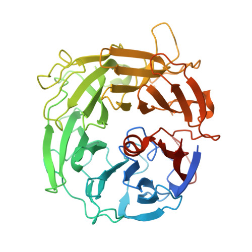
The human gut microbiota use complex carbohydrates as major nutrients. The requirement for an efficient glycan degrading systems exerts a major selection pressure on this microbial community. Thus, we propose that these bacteria represent a substantial resource for discovering novel carbohydrate active enzymes. To test this hypothesis, we focused on enzymes that hydrolyze rhamnosidic bonds, as cleavage of these linkages is chemically challenging and there is a paucity of information on l-rhamnosidases. Here we screened the activity of enzymes derived from the human gut microbiota bacterium Bacteroides thetaiotaomicron , which are up-regulated in response to rhamnose-containing glycans. We identified an α-l-rhamnosidase, BT3686, which is the founding member of a glycoside hydrolase (GH) family, GH145. In contrast to other rhamnosidases, BT3686 cleaved l-Rha-α1,4-d-GlcA linkages through a retaining double-displacement mechanism. The crystal structure of BT3686 showed that the enzyme displayed a type A seven-bladed β-propeller fold. Mutagenesis and crystallographic studies, including the structure of BT3686 in complex with the reaction product GlcA, revealed a location for the active site among β-propeller enzymes cited on the posterior surface of the rhamnosidase. In contrast to the vast majority of GH, the catalytic apparatus of BT3686 does not comprise a pair of carboxylic acid residues but, uniquely, a single histidine functions as the only discernable catalytic amino acid. Intriguingly, the histidine, His48, is not invariant in GH145; however, when engineered into structural homologs lacking the imidazole residue, α-l-rhamnosidase activity was established. The potential contribution of His48 to the catalytic activity of BT3686 is discussed.
- Institute for Cell and Molecular Biosciences, Newcastle University, Newcastle upon Tyne NE2 4HH, United Kingdom.
Organizational Affiliation: