5MUA | pdb_00005mua

PSL1a-E64 complex


Experimental Data Snapshot

  • Method: X-RAY DIFFRACTION
  • Resolution: 1.49 Å
  • R-Value Free: 
    0.248 (Depositor), 0.249 (DCC) 
  • R-Value Work: 
    0.221 (Depositor), 0.224 (DCC) 
  • R-Value Observed: 
    0.222 (Depositor) 

Starting Model: experimental
View more details

wwPDB Validation 3D Report Full Report

Validation slider image for 5MUA

Ligand Structure Quality Assessment 


This is version 1.5 of the entry. See complete history

Literature

Family of Papain-Like Fungal Chimerolectins with Distinct Ca(2+)-Dependent Activation Mechanism.

Cordara, G.Manna, D.Krengel, U.

(2017) Biochemistry 56: 4689-4700

  • DOI: https://doi.org/10.1021/acs.biochem.7b00317
  • Primary Citation Related Structures: 
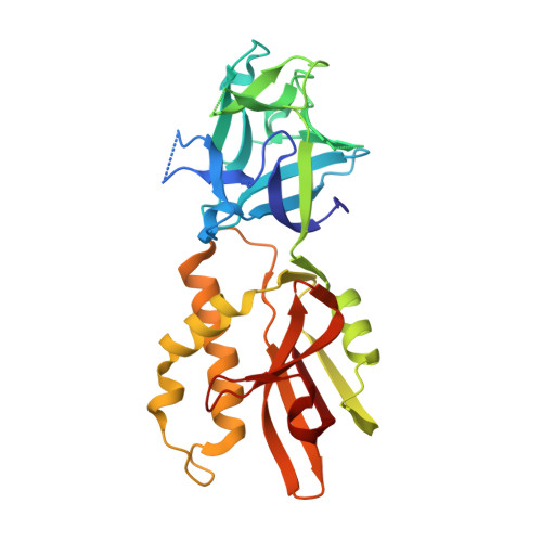
    5MU9, 5MUA

  • PubMed Abstract: 

    An important function of fungal lectins is to protect their host. Marasmius oreades agglutinin (MOA) is toxic to nematodes and exerts its protective effect through protease activity. Its proteolytic function is associated with a papain-like dimerization domain. The closest homologue of MOA is Polyporus squamosus lectin 1a (PSL1a). Here, we probed PSL1a for catalytic activity and confirmed that it is a calcium-dependent cysteine protease, like MOA. The X-ray crystal structures of PSL1a (1.5 Å) and MOA (1.3 Å) in complex with calcium and the irreversible cysteine protease inhibitor E-64 elucidated the structural basis for their mechanism of action. The comparison with other calcium-dependent proteases (calpains, LapG) reveals a unique metal-dependent activation mechanism relying on a calcium-induced backbone shift and intradimer cooperation. Intriguingly, the enzymes appear to use a tyrosine-gating mechanism instead of pro-peptide processing. A search for potential MOA orthologues suggests the existence of a whole new family of fungal chimerolectins with these unique features.


  • Organizational Affiliation
    • Department of Chemistry, University of Oslo , P.O. Box 1033, Blindern, 0315 Oslo, Norway.

Macromolecule Content 

  • Total Structure Weight: 63.75 kDa 
  • Atom Count: 4,925 
  • Modeled Residue Count: 544 
  • Deposited Residue Count: 572 
  • Unique protein chains: 1

Macromolecules

Find similar proteins by:|  3D Structure
Entity ID: 1
MoleculeChains  Sequence LengthOrganismDetailsImage
Ricin B-related lectin
A, B
286Cerioporus squamosusMutation(s): 0 
Gene Names: psl1a
UniProt
Find proteins for Q75WT9 (Cerioporus squamosus)
Explore Q75WT9 
Go to UniProtKB:  Q75WT9
Entity Groups
Sequence Clusters30% Identity50% Identity70% Identity90% Identity95% Identity100% Identity
UniProt GroupQ75WT9
Sequence Annotations
Expand
Reference Sequence

Small Molecules

Ligands 4 Unique
IDChains Name / Formula / InChI Key2D Diagram3D Interactions
E64

Query on E64


Download:Ideal Coordinates CCD File
F [auth A],
K [auth B]
N-[N-[1-HYDROXYCARBOXYETHYL-CARBONYL]LEUCYLAMINO-BUTYL]-GUANIDINE
C15 H30 N5 O5
QPQNJAXBPHVASB-QWRGUYRKSA-O
GAL

Query on GAL


Download:Ideal Coordinates CCD File
C [auth A],
J [auth B]
beta-D-galactopyranose
C6 H12 O6
WQZGKKKJIJFFOK-FPRJBGLDSA-N
DMS

Query on DMS


Download:Ideal Coordinates CCD File
G [auth A]DIMETHYL SULFOXIDE
C2 H6 O S
IAZDPXIOMUYVGZ-UHFFFAOYSA-N
CA

Query on CA


Download:Ideal Coordinates CCD File
D [auth A],
E [auth A],
H [auth A],
I [auth A]
CALCIUM ION
Ca
BHPQYMZQTOCNFJ-UHFFFAOYSA-N

Experimental Data & Validation

Experimental Data

  • Method: X-RAY DIFFRACTION
  • Resolution: 1.49 Å
  • R-Value Free:  0.248 (Depositor), 0.249 (DCC) 
  • R-Value Work:  0.221 (Depositor), 0.224 (DCC) 
  • R-Value Observed: 0.222 (Depositor) 
Space Group: P 21 21 21
Unit Cell:
Length ( Å )Angle ( ˚ )
a = 75.671α = 90
b = 94.331β = 90
c = 102.764γ = 90
Software Package:
Software NamePurpose
REFMACrefinement
XDSdata reduction
XDSdata scaling
PHASERphasing
Cootmodel building

Structure Validation

View Full Validation Report


Ligand Structure Quality Assessment 


Entry History 

Deposition Data

Revision History  (Full details and data files)

  • Version 1.0: 2017-07-12
    Type: Initial release
  • Version 1.1: 2017-08-30
    Changes: Database references
  • Version 1.2: 2017-09-13
    Changes: Database references
  • Version 1.3: 2020-07-29
    Type: Remediation
    Reason: Carbohydrate remediation
    Changes: Data collection, Derived calculations, Structure summary
  • Version 1.4: 2024-01-17
    Changes: Data collection, Database references, Refinement description, Structure summary
  • Version 1.5: 2024-11-20
    Changes: Structure summary