


Crystal structure of human carbonic anhydrase isozyme XII with 2,3,5,6-Tetrafluoro-4-(propylthio)benzenesulfonamide
Smirnov, A., Manakova, E., Grazulis, S., Matulis, D., Dudutiene, V.To be published.
Experimental Data Snapshot
Starting Model: experimental
View more details
Entity ID: 1 | |||||
|---|---|---|---|---|---|
| Molecule | Chains | Sequence Length | Organism | Details | Image |
| Carbonic anhydrase 12 | 263 | Homo sapiens | Mutation(s): 0 Gene Names: CA12 EC: 4.2.1.1 | 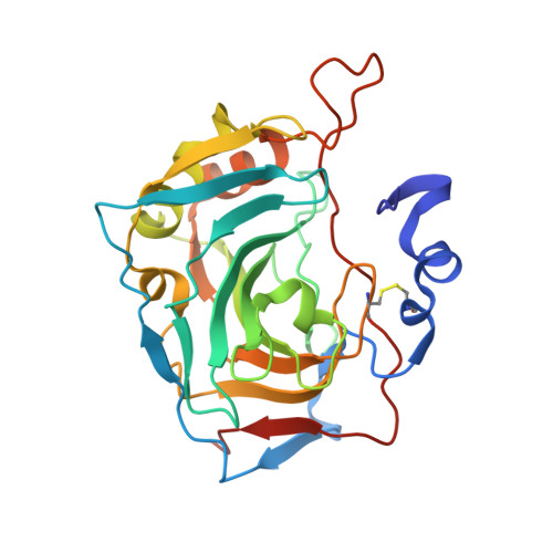 | |
UniProt & NIH Common Fund Data Resources | |||||
PHAROS: O43570 GTEx: ENSG00000074410 | |||||
Entity Groups | |||||
| Sequence Clusters | 30% Identity50% Identity70% Identity90% Identity95% Identity100% Identity | ||||
| UniProt Group | O43570 | ||||
Sequence AnnotationsExpand | |||||
Reference Sequence | |||||
| Ligands 5 Unique | |||||
|---|---|---|---|---|---|
| ID | Chains | Name / Formula / InChI Key | 2D Diagram | 3D Interactions | |
| 3TV Download:Ideal Coordinates CCD File | F [auth A], P [auth B], U [auth C], Z [auth D] | 2,3,5,6-tetrafluoro-4-(propylsulfanyl)benzenesulfonamide C9 H9 F4 N O2 S2 LKPIFWBRFVJTMY-UHFFFAOYSA-N |  | ||
| CIT Download:Ideal Coordinates CCD File | BA [auth D], DA [auth D], S [auth B] | CITRIC ACID C6 H8 O7 KRKNYBCHXYNGOX-UHFFFAOYSA-N |  | ||
| PEG Download:Ideal Coordinates CCD File | AA [auth D], J [auth A], R [auth B], W [auth C] | DI(HYDROXYETHYL)ETHER C4 H10 O3 MTHSVFCYNBDYFN-UHFFFAOYSA-N |  | ||
| ZN Download:Ideal Coordinates CCD File | E [auth A], O [auth B], T [auth C], Y [auth D] | ZINC ION Zn PTFCDOFLOPIGGS-UHFFFAOYSA-N |  | ||
| EDO Download:Ideal Coordinates CCD File | CA [auth D] EA [auth D] G [auth A] H [auth A] I [auth A] | 1,2-ETHANEDIOL C2 H6 O2 LYCAIKOWRPUZTN-UHFFFAOYSA-N |  | ||
| Length ( Å ) | Angle ( ˚ ) |
|---|---|
| a = 77.17 | α = 90 |
| b = 74.157 | β = 108.85 |
| c = 91.559 | γ = 90 |
| Software Name | Purpose |
|---|---|
| REFMAC | refinement |
| SCALA | data scaling |
| PDB_EXTRACT | data extraction |
| XDS | data reduction |
| MOLREP | phasing |


