5MR6 | pdb_00005mr6

XiaF from Streptomyces sp. in complex with FADH2 and Glycerol


Experimental Data Snapshot

  • Method: X-RAY DIFFRACTION
  • Resolution: 2.40 Å
  • R-Value Free: 
    0.229 (Depositor), 0.186 (DCC) 
  • R-Value Work: 
    0.198 (Depositor) 
  • R-Value Observed: 
    0.200 (Depositor) 

Starting Model: experimental
View more details

wwPDB Validation 3D Report Full Report

Validation slider image for 5MR6

Ligand Structure Quality Assessment 


This is version 1.2 of the entry. See complete history

Literature

Cryptic indole hydroxylation by a non-canonical terpenoid cyclase parallels bacterial xenobiotic detoxification.

Kugel, S.Baunach, M.Baer, P.Ishida-Ito, M.Sundaram, S.Xu, Z.Groll, M.Hertweck, C.

(2017) Nat Commun 8: 15804-15804

  • DOI: https://doi.org/10.1038/ncomms15804
  • Primary Citation Related Structures: 
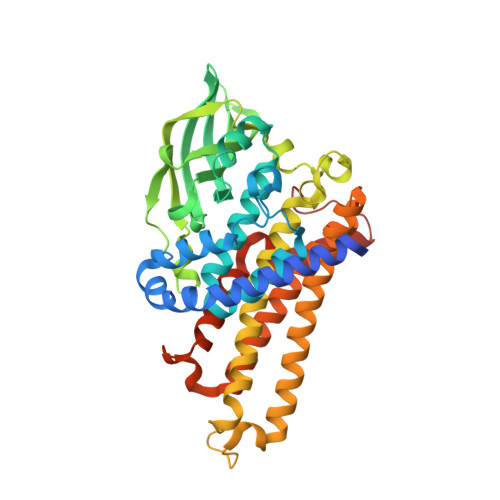
    5LVU, 5LVW, 5MR6

  • PubMed Abstract: 

    Terpenoid natural products comprise a wide range of molecular architectures that typically result from C-C bond formations catalysed by classical type I/II terpene cyclases. However, the molecular diversity of biologically active terpenoids is substantially increased by fully unrelated, non-canonical terpenoid cyclases. Their evolutionary origin has remained enigmatic. Here we report the in vitro reconstitution of an unusual flavin-dependent bacterial indoloterpenoid cyclase, XiaF, together with a designated flavoenzyme-reductase (XiaP) that mediates a key step in xiamycin biosynthesis. The crystal structure of XiaF with bound FADH 2 (at 2.4 Å resolution) and phylogenetic analyses reveal that XiaF is, surprisingly, most closely related to xenobiotic-degrading enzymes. Biotransformation assays show that XiaF is a designated indole hydroxylase that can be used for the production of indigo and indirubin. We unveil a cryptic hydroxylation step that sets the basis for terpenoid cyclization and suggest that the cyclase has evolved from xenobiotics detoxification enzymes.


  • Organizational Affiliation
    • Department of Biomolecular Chemistry, Leibniz Institute for Natural Product Research and Infection Biology (HKI), Beutenbergstr. 11a, 07745 Jena, Germany.

Macromolecule Content 

  • Total Structure Weight: 1,082.82 kDa 
  • Atom Count: 77,543 
  • Modeled Residue Count: 9,504 
  • Deposited Residue Count: 9,912 
  • Unique protein chains: 1

Macromolecules

Find similar proteins by:|  3D Structure
Entity ID: 1
MoleculeChains  Sequence LengthOrganismDetailsImage
XiaF protein
A, B, C, D, E
A, B, C, D, E, F, G, H, I, J, K, L, M, N, O, P, Q, R, S, T, U, V, W, X
413Streptomyces sp.Mutation(s): 0 
Gene Names: xiaF
UniProt
Find proteins for I7IIA9 (Streptomyces sp. HKI0576)
Explore I7IIA9 
Go to UniProtKB:  I7IIA9
Entity Groups
Sequence Clusters30% Identity50% Identity70% Identity90% Identity95% Identity100% Identity
UniProt GroupI7IIA9
Sequence Annotations
Expand
Reference Sequence

Small Molecules

Ligands 2 Unique
IDChains Name / Formula / InChI Key2D Diagram3D Interactions
FAD

Query on FAD


Download:Ideal Coordinates CCD File
AA [auth B]
AB [auth O]
CA [auth C]
CB [auth P]
EA [auth D]
AA [auth B],
AB [auth O],
CA [auth C],
CB [auth P],
EA [auth D],
EB [auth Q],
GA [auth E],
GB [auth R],
IA [auth F],
IB [auth S],
KA [auth G],
KB [auth T],
MA [auth H],
MB [auth U],
OA [auth I],
OB [auth V],
QA [auth J],
QB [auth W],
SA [auth K],
SB [auth X],
UA [auth L],
WA [auth M],
Y [auth A],
YA [auth N]
FLAVIN-ADENINE DINUCLEOTIDE
C27 H33 N9 O15 P2
VWWQXMAJTJZDQX-UYBVJOGSSA-N
GOL

Query on GOL


Download:Ideal Coordinates CCD File
BA [auth B]
BB [auth O]
DA [auth C]
DB [auth P]
FA [auth D]
BA [auth B],
BB [auth O],
DA [auth C],
DB [auth P],
FA [auth D],
FB [auth Q],
HA [auth E],
HB [auth R],
JA [auth F],
JB [auth S],
LA [auth G],
LB [auth T],
NA [auth H],
NB [auth U],
PA [auth I],
PB [auth V],
RA [auth J],
RB [auth W],
TA [auth K],
TB [auth X],
VA [auth L],
XA [auth M],
Z [auth A],
ZA [auth N]
GLYCEROL
C3 H8 O3
PEDCQBHIVMGVHV-UHFFFAOYSA-N

Experimental Data & Validation

Experimental Data

  • Method: X-RAY DIFFRACTION
  • Resolution: 2.40 Å
  • R-Value Free:  0.229 (Depositor), 0.186 (DCC) 
  • R-Value Work:  0.198 (Depositor) 
  • R-Value Observed: 0.200 (Depositor) 
Space Group: P 1
Unit Cell:
Length ( Å )Angle ( ˚ )
a = 166.93α = 89.96
b = 167.09β = 90.18
c = 200.94γ = 119.82
Software Package:
Software NamePurpose
REFMACrefinement
XDSdata reduction
XSCALEdata scaling
PHASERphasing

Structure Validation

View Full Validation Report


Ligand Structure Quality Assessment 


Entry History 

& Funding Information

Deposition Data


Funding OrganizationLocationGrant Number
German Research FoundationGermanySFB749

Revision History  (Full details and data files)

  • Version 1.0: 2017-06-28
    Type: Initial release
  • Version 1.1: 2017-07-05
    Changes: Database references
  • Version 1.2: 2024-01-17
    Changes: Author supporting evidence, Data collection, Database references, Refinement description