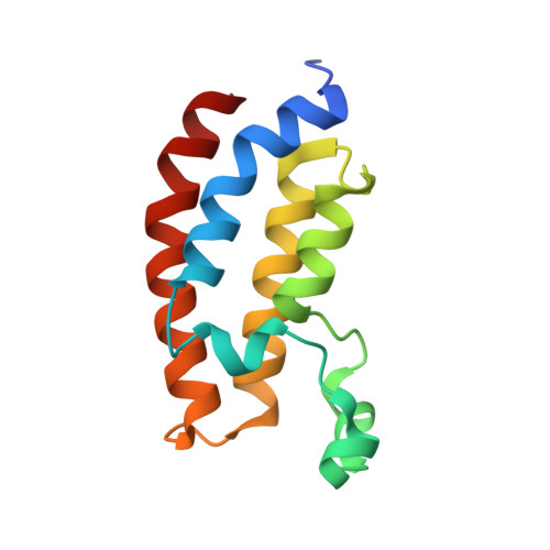
5MQG

Crystal structure of CREBBP bromodomain complexed with CBP015


Experimental Data Snapshot

  • Method: X-RAY DIFFRACTION
  • Resolution: 1.35 Å
  • R-Value Free: 0.191 
  • R-Value Work: 0.171 
  • R-Value Observed: 0.172 

wwPDB Validation   3D Report Full Report


Ligand Structure Quality Assessment 


This is version 1.5 of the entry. See complete history


Literature

Virtual screen to NMR (VS2NMR): Discovery of fragment hits for the CBP bromodomain.

Spiliotopoulos, D.Zhu, J.Wamhoff, E.C.Deerain, N.Marchand, J.R.Aretz, J.Rademacher, C.Caflisch, A.

(2017) Bioorg Med Chem Lett 27: 2472-2478

  • DOI: https://doi.org/10.1016/j.bmcl.2017.04.001
  • Primary Citation of Related Structures:  
    5MPZ, 5MQE, 5MQG, 5MQK

  • PubMed Abstract: 

    Overexpression of the CREB-binding protein (CBP), a bromodomain-containing transcription coactivator involved in a variety of cellular processes, has been observed in several types of cancer with a correlation to aggressiveness. We have screened a library of nearly 1500 fragments by high-throughput docking into the CBP bromodomain followed by binding energy evaluation using a force field with electrostatic solvation. Twenty of the 39 fragments selected by virtual screening are positive in one or more ligand-observed nuclear magnetic resonance (NMR) experiments. Four crystal structures of the CBP bromodomain in complex with in silico screening hits validate the pose predicted by docking. Thus, the success ratio of the high-throughput docking procedure is 50% or 10% if one considers the validation by ligand-observed NMR spectroscopy or X-ray crystallography, respectively. Compounds 1 and 3 show favorable ligand efficiency in two different in vitro binding assays. The structure of the CBP bromodomain in the complex with the brominated pyrrole 1 suggests fragment growing by Suzuki coupling.


  • Organizational Affiliation

    Department of Biochemistry, University of Zürich, Winterthurerstrasse 190, CH-8057 Zürich, Switzerland. Electronic address: d.spiliotopoulos@bioc.uzh.ch.


Macromolecules
Find similar proteins by:  (by identity cutoff)  |  3D Structure
Entity ID: 1
MoleculeChains Sequence LengthOrganismDetailsImage
CREB-binding protein
A, B
119Homo sapiensMutation(s): 0 
Gene Names: CREBBPCBP
EC: 2.3.1.48 (PDB Primary Data), 2.3.1 (UniProt)
UniProt & NIH Common Fund Data Resources
Find proteins for Q92793 (Homo sapiens)
Explore Q92793 
Go to UniProtKB:  Q92793
PHAROS:  Q92793
GTEx:  ENSG00000005339 
Entity Groups  
Sequence Clusters30% Identity50% Identity70% Identity90% Identity95% Identity100% Identity
UniProt GroupQ92793
Sequence Annotations
Expand
  • Reference Sequence
Small Molecules
Ligands 1 Unique
IDChains Name / Formula / InChI Key2D Diagram3D Interactions
F31
Query on F31

Download Ideal Coordinates CCD File 
C [auth A],
D [auth B]
1-(4-azanyl-3-methoxy-phenyl)ethanone
C9 H11 N O2
LNRPHCWGORQWPI-UHFFFAOYSA-N
Experimental Data & Validation

Experimental Data

  • Method: X-RAY DIFFRACTION
  • Resolution: 1.35 Å
  • R-Value Free: 0.191 
  • R-Value Work: 0.171 
  • R-Value Observed: 0.172 
  • Space Group: H 3
Unit Cell:
Length ( Å )Angle ( ˚ )
a = 121.436α = 90
b = 121.436β = 90
c = 40.271γ = 120
Software Package:
Software NamePurpose
PHENIXrefinement
XDSdata reduction
SCALAdata scaling
PHASERphasing

Structure Validation

View Full Validation Report


Ligand Structure Quality Assessment 


Entry History 

Deposition Data

Revision History  (Full details and data files)

  • Version 1.0: 2017-04-19
    Type: Initial release
  • Version 1.1: 2017-04-26
    Changes: Database references
  • Version 1.2: 2017-05-10
    Changes: Structure summary
  • Version 1.3: 2017-05-17
    Changes: Database references
  • Version 1.4: 2019-10-16
    Changes: Data collection
  • Version 1.5: 2024-05-08
    Changes: Data collection, Database references