5MJD

metNgb under oxygen at 80 bar


Experimental Data Snapshot

  • Method: X-RAY DIFFRACTION
  • Resolution: 1.70 Å
  • R-Value Free: 0.242 
  • R-Value Work: 0.179 
  • R-Value Observed: 0.182 

Starting Model: experimental
View more details

wwPDB Validation   3D Report Full Report


Ligand Structure Quality Assessment 


This is version 1.5 of the entry. See complete history


Literature

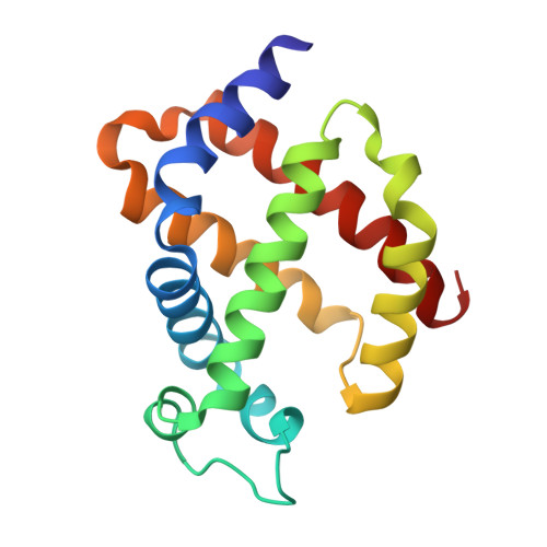
Ligand pathways in neuroglobin revealed by low-temperature photodissociation and docking experiments.

Ardiccioni, C.Arcovito, A.Della Longa, S.van der Linden, P.Bourgeois, D.Weik, M.Montemiglio, L.C.Savino, C.Avella, G.Exertier, C.Carpentier, P.Prange, T.Brunori, M.Colloc'h, N.Vallone, B.

(2019) IUCrJ 6: 832-842

  • DOI: https://doi.org/10.1107/S2052252519008157
  • Primary Citation of Related Structures:  
    5MJC, 5MJD, 6I3T, 6I40

  • PubMed Abstract: 

    A combined biophysical approach was applied to map gas-docking sites within murine neuroglobin (Ngb), revealing snapshots of events that might govern activity and dynamics in this unique hexacoordinate globin, which is most likely to be involved in gas-sensing in the central nervous system and for which a precise mechanism of action remains to be elucidated. The application of UV-visible microspectroscopy in crystallo , solution X-ray absorption near-edge spectroscopy and X-ray diffraction experiments at 15-40 K provided the structural characterization of an Ngb photolytic intermediate by cryo-trapping and allowed direct observation of the relocation of carbon monoxide within the distal heme pocket after photodissociation. Moreover, X-ray diffraction at 100 K under a high pressure of dioxygen, a physiological ligand of Ngb, unravelled the existence of a storage site for O 2 in Ngb which coincides with Xe-III, a previously described docking site for xenon or krypton. Notably, no other secondary sites were observed under our experimental conditions.


  • Organizational Affiliation

    Department of Life and Environmental Sciences, New York-Marche Structural Biology Center (NY-MaSBiC), Polytechnic University of Marche, Ancona, Italy.


Macromolecules
Find similar proteins by:  (by identity cutoff)  |  3D Structure
Entity ID: 1
MoleculeChains Sequence LengthOrganismDetailsImage
Neuroglobin148Mus musculusMutation(s): 2 
Gene Names: Ngb
UniProt
Find proteins for Q9ER97 (Mus musculus)
Explore Q9ER97 
Go to UniProtKB:  Q9ER97
Entity Groups  
Sequence Clusters30% Identity50% Identity70% Identity90% Identity95% Identity100% Identity
UniProt GroupQ9ER97
Sequence Annotations
Expand
  • Reference Sequence
Experimental Data & Validation

Experimental Data

  • Method: X-RAY DIFFRACTION
  • Resolution: 1.70 Å
  • R-Value Free: 0.242 
  • R-Value Work: 0.179 
  • R-Value Observed: 0.182 
  • Space Group: H 3 2
Unit Cell:
Length ( Å )Angle ( ˚ )
a = 87.819α = 90
b = 87.819β = 90
c = 113.522γ = 120
Software Package:
Software NamePurpose
REFMACrefinement
XDSdata reduction
XSCALEdata scaling
REFMACphasing

Structure Validation

View Full Validation Report


Ligand Structure Quality Assessment 


Entry History 

Deposition Data

Revision History  (Full details and data files)

  • Version 1.0: 2017-12-20
    Type: Initial release
  • Version 1.1: 2018-02-21
    Changes: Database references, Structure summary
  • Version 1.2: 2019-07-10
    Changes: Data collection, Database references, Structure summary
  • Version 1.3: 2019-09-11
    Changes: Data collection, Database references
  • Version 1.4: 2019-10-09
    Changes: Data collection, Database references
  • Version 1.5: 2024-01-17
    Changes: Data collection, Database references, Refinement description