5MIM | pdb_00005mim

Xray structure of human furin bound with the 2,5-dideoxystreptamine derived small molecule inhibitor 1n


Experimental Data Snapshot

  • Method: X-RAY DIFFRACTION
  • Resolution: 1.90 Å
  • R-Value Free: 
    0.184 (Depositor), 0.185 (DCC) 
  • R-Value Work: 
    0.165 (Depositor), 0.167 (DCC) 
  • R-Value Observed: 
    0.166 (Depositor) 

Starting Model: experimental
View more details

wwPDB Validation 3D Report Full Report

Validation slider image for 5MIM

Ligand Structure Quality Assessment 


This is version 1.3 of the entry. See complete history

Literature

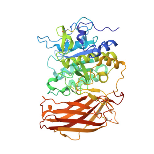
Structural Studies Revealed Active Site Distortions of Human Furin by a Small Molecule Inhibitor.

Dahms, S.O.Jiao, G.S.Than, M.E.

(2017) ACS Chem Biol 12: 1211-1216

  • DOI: https://doi.org/10.1021/acschembio.6b01110
  • Primary Citation Related Structures: 
    5MIM

  • PubMed Abstract: 

    Proprotein convertases (PCs) represent highly selective serine proteases that activate their substrates upon proteolytic cleavage. Their inhibition is a promising strategy for the treatment of several pathologies including cancer, atherosclerosis, hypercholesterolaemia, and infectious diseases. Here, we present the first experimental complex of furin with a non-substrate-like small molecule inhibitor, and the X-ray structure of the enzyme complexed to the small molecule inhibitor 1 at 1.9 Å resolution. Two molecules of inhibitor 1 were found to interact with furin. One is anchored at the S4 pocket of the enzyme and interferes directly with the conformation and function of the catalytic triade; the other molecule shows weaker binding and interacts with a distant, less conserved region of furin. The observed binding modes represent a new inhibition strategy of furin and imply the possibility to attain specificity among the PCs providing an innovative starting point of structure guided inhibitor development for furin.


  • Organizational Affiliation
    • Protein Crystallography Group, Leibniz Institute on Aging - Fritz Lipmann Institute (FLI) , Beutenbergstr. 11, 07745 Jena, Germany.

Macromolecule Content 

  • Total Structure Weight: 53.81 kDa 
  • Atom Count: 4,275 
  • Modeled Residue Count: 473 
  • Deposited Residue Count: 482 
  • Unique protein chains: 1

Macromolecules

Find similar proteins by:|  3D Structure
Entity ID: 1
MoleculeChains  Sequence LengthOrganismDetailsImage
Furin482Homo sapiensMutation(s): 0 
Gene Names: FURINFURPACEPCSK3
EC: 3.4.21.75
UniProt & NIH Common Fund Data Resources
Find proteins for P09958 (Homo sapiens)
Explore P09958 
Go to UniProtKB:  P09958
PHAROS:  P09958
GTEx:  ENSG00000140564 
Entity Groups
Sequence Clusters30% Identity50% Identity70% Identity90% Identity95% Identity100% Identity
UniProt GroupP09958
Sequence Annotations
Expand
Reference Sequence

Small Molecules

Ligands 4 Unique
IDChains Name / Formula / InChI Key2D Diagram3D Interactions
1N

Query on 1N


Download:Ideal Coordinates CCD File
J [auth A],
K [auth A]
1-[(1~{R},2~{R},4~{S},5~{S})-2,4-bis(4-carbamimidamidophenoxy)-5-[(4-carbamimidamidophenyl)amino]cyclohexyl]guanidine
C28 H37 N13 O2
PKMXMBXOZUEHDV-UARRHKHWSA-N
CA

Query on CA


Download:Ideal Coordinates CCD File
B [auth A],
C [auth A],
D [auth A]
CALCIUM ION
Ca
BHPQYMZQTOCNFJ-UHFFFAOYSA-N
CL

Query on CL


Download:Ideal Coordinates CCD File
I [auth A]CHLORIDE ION
Cl
VEXZGXHMUGYJMC-UHFFFAOYSA-M
NA

Query on NA


Download:Ideal Coordinates CCD File
E [auth A],
F [auth A],
G [auth A],
H [auth A]
SODIUM ION
Na
FKNQFGJONOIPTF-UHFFFAOYSA-N

Experimental Data & Validation

Experimental Data

  • Method: X-RAY DIFFRACTION
  • Resolution: 1.90 Å
  • R-Value Free:  0.184 (Depositor), 0.185 (DCC) 
  • R-Value Work:  0.165 (Depositor), 0.167 (DCC) 
  • R-Value Observed: 0.166 (Depositor) 
Space Group: P 65 2 2
Unit Cell:
Length ( Å )Angle ( ˚ )
a = 132.204α = 90
b = 132.204β = 90
c = 155.727γ = 120
Software Package:
Software NamePurpose
PHENIXrefinement
XDSdata reduction
XDSdata scaling
PHENIXphasing

Structure Validation

View Full Validation Report


Ligand Structure Quality Assessment 


Entry History 

Deposition Data

Revision History  (Full details and data files)

  • Version 1.0: 2017-05-10
    Type: Initial release
  • Version 1.1: 2017-05-31
    Changes: Database references
  • Version 1.2: 2024-01-17
    Changes: Advisory, Data collection, Database references, Derived calculations, Refinement description
  • Version 1.3: 2024-11-20
    Changes: Structure summary