5MIJ | pdb_00005mij

X-ray structure of carboplatin-encapsulated horse spleen apoferritin


Experimental Data Snapshot

  • Method: X-RAY DIFFRACTION
  • Resolution: 1.49 Å
  • R-Value Free: 
    0.181 (Depositor), 0.184 (DCC) 
  • R-Value Work: 
    0.158 (Depositor), 0.162 (DCC) 
  • R-Value Observed: 
    0.159 (Depositor) 

wwPDB Validation 3D Report Full Report

Validation slider image for 5MIJ

This is version 1.2 of the entry. See complete history

Literature

X-ray Structure of the Carboplatin-Loaded Apo-Ferritin Nanocage.

Pontillo, N.Ferraro, G.Helliwell, J.R.Amoresano, A.Merlino, A.

(2017) ACS Med Chem Lett 8: 433-437

  • DOI: https://doi.org/10.1021/acsmedchemlett.7b00025
  • Primary Citation Related Structures: 
    5MIJ, 5MIK

  • PubMed Abstract: 

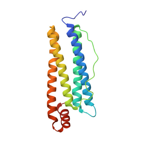
    The second-generation Pt anticancer agent carboplatin (CBDCA) was encapsulated within the apo horse spleen ferritin (AFt) nanocage, and the X-ray structure of the drug-loaded protein was refined at 1.49 Å resolution. Two Pt binding sites, different from the one observed in the cisplatin-encapsulated AFt, were identified in Ft subunits by inspection of anomalous electron density maps at two wavelengths and difference Fourier electron density maps, which provide the necessary sensitivity to discriminate between Pt from CBDCA and Cd ions that are present in the crystallization conditions. Pt centers coordinate to the NE2 atom of His49 and to the NE2 atom of His132, both on the inner surface of the Ft nanocage.


  • Organizational Affiliation
    • Department of Chemical Sciences, University of Naples Federico II, Complesso Universitario di Monte Sant'Angelo, Via Cintia, I-80126 Napoli, Italy.

Macromolecule Content 

  • Total Structure Weight: 21.74 kDa 
  • Atom Count: 1,782 
  • Modeled Residue Count: 172 
  • Deposited Residue Count: 174 
  • Unique protein chains: 1

Macromolecules

Find similar proteins by:|  3D Structure
Entity ID: 1
MoleculeChains  Sequence LengthOrganismDetailsImage
Ferritin light chain174Equus caballusMutation(s): 0 
UniProt
Find proteins for P02791 (Equus caballus)
Explore P02791 
Go to UniProtKB:  P02791
Entity Groups
Sequence Clusters30% Identity50% Identity70% Identity90% Identity95% Identity100% Identity
UniProt GroupP02791
Sequence Annotations
Expand
Reference Sequence

Small Molecules

Ligands 4 Unique
IDChains Name / Formula / InChI Key2D Diagram3D Interactions
PT

Query on PT


Download:Ideal Coordinates CCD File
Q [auth A],
R [auth A]
PLATINUM (II) ION
Pt
HRGDZIGMBDGFTC-UHFFFAOYSA-N
CD

Query on CD


Download:Ideal Coordinates CCD File
B [auth A]
C [auth A]
D [auth A]
E [auth A]
F [auth A]
B [auth A],
C [auth A],
D [auth A],
E [auth A],
F [auth A],
G [auth A],
H [auth A],
I [auth A],
J [auth A],
K [auth A],
O [auth A]
CADMIUM ION
Cd
WLZRMCYVCSSEQC-UHFFFAOYSA-N
SO4

Query on SO4


Download:Ideal Coordinates CCD File
P [auth A]SULFATE ION
O4 S
QAOWNCQODCNURD-UHFFFAOYSA-L
CL

Query on CL


Download:Ideal Coordinates CCD File
L [auth A],
M [auth A],
N [auth A],
S [auth A]
CHLORIDE ION
Cl
VEXZGXHMUGYJMC-UHFFFAOYSA-M

Experimental Data & Validation

Experimental Data

  • Method: X-RAY DIFFRACTION
  • Resolution: 1.49 Å
  • R-Value Free:  0.181 (Depositor), 0.184 (DCC) 
  • R-Value Work:  0.158 (Depositor), 0.162 (DCC) 
  • R-Value Observed: 0.159 (Depositor) 
Space Group: F 4 3 2
Unit Cell:
Length ( Å )Angle ( ˚ )
a = 181.67α = 90
b = 181.67β = 90
c = 181.67γ = 90
Software Package:
Software NamePurpose
REFMACrefinement
iMOSFLMdata reduction
SCALAdata scaling
PHASERphasing

Structure Validation

View Full Validation Report


Entry History 

Deposition Data

Revision History  (Full details and data files)

  • Version 1.0: 2017-03-15
    Type: Initial release
  • Version 1.1: 2017-05-03
    Changes: Database references
  • Version 1.2: 2024-05-08
    Changes: Data collection, Database references, Derived calculations