5MFP | pdb_00005mfp

Human Sirt6 in complex with activator UBCS58


Experimental Data Snapshot

  • Method: X-RAY DIFFRACTION
  • Resolution: 1.98 Å
  • R-Value Free: 
    0.185 (Depositor), 0.180 (DCC) 
  • R-Value Work: 
    0.157 (Depositor) 
  • R-Value Observed: 
    0.159 (Depositor) 

Starting Model: experimental
View more details

wwPDB Validation 3D Report Full Report

Validation slider image for 5MFP

Ligand Structure Quality Assessment 


This is version 1.2 of the entry. See complete history

Literature

Structural Basis of Sirtuin 6 Activation by Synthetic Small Molecules.

You, W.Rotili, D.Li, T.M.Kambach, C.Meleshin, M.Schutkowski, M.Chua, K.F.Mai, A.Steegborn, C.

(2017) Angew Chem Int Ed Engl 56: 1007-1011

  • DOI: https://doi.org/10.1002/anie.201610082
  • Primary Citation Related Structures: 
    5MF6, 5MFP, 5MFZ, 5MGN

  • PubMed Abstract: 

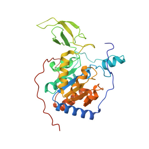
    Sirtuins are protein deacylases regulating metabolism and stress responses, and are implicated in aging-related diseases. Small molecule activators for the human sirtuins Sirt1-7 are sought as chemical tools and potential therapeutics, such as for cancer. Activators are available for Sirt1 and exploit its unique N-terminus, whereas drug-like activators for Sirt2-7 are lacking. We synthesized and screened pyrrolo[1,2-a]quinoxaline derivatives, yielding the first synthetic Sirt6 activators. Biochemical assays show direct, substrate-independent compound binding to the Sirt6 catalytic core and potent activation of Sirt6-dependent deacetylation of peptide substrates and complete nucleosomes. Crystal structures of Sirt6/activator complexes reveal that the compounds bind to a Sirt6-specific acyl channel pocket and identify key interactions. Our results establish potent Sirt6 activation with small molecules and provide a structural basis for further development of Sirt6 activators as tools and therapeutics.


  • Organizational Affiliation
    • Universität Bayreuth, Lehrstuhl Biochemie und Forschungszentrum für Biomakromoleküle, Universitätsstr. 30, 95447, Bayreuth, Germany.

Macromolecule Content 

  • Total Structure Weight: 70.46 kDa 
  • Atom Count: 4,719 
  • Modeled Residue Count: 557 
  • Deposited Residue Count: 604 
  • Unique protein chains: 1

Macromolecules

Find similar proteins by:|  3D Structure
Entity ID: 1
MoleculeChains  Sequence LengthOrganismDetailsImage
NAD-dependent protein deacetylase sirtuin-6
A, B
302Homo sapiensMutation(s): 0 
Gene Names: SIRT6SIR2L6
EC: 3.5.1 (PDB Primary Data), 2.4.2 (UniProt), 2.3.1 (UniProt), 2.3.1.286 (UniProt)
UniProt & NIH Common Fund Data Resources
Find proteins for Q8N6T7 (Homo sapiens)
Explore Q8N6T7 
Go to UniProtKB:  Q8N6T7
PHAROS:  Q8N6T7
GTEx:  ENSG00000077463 
Entity Groups
Sequence Clusters30% Identity50% Identity70% Identity90% Identity95% Identity100% Identity
UniProt GroupQ8N6T7
Sequence Annotations
Expand
Reference Sequence

Small Molecules

Ligands 7 Unique
IDChains Name / Formula / InChI Key2D Diagram3D Interactions
AR6

Query on AR6


Download:Ideal Coordinates CCD File
C [auth A],
N [auth B]
[(2R,3S,4R,5R)-5-(6-AMINOPURIN-9-YL)-3,4-DIHYDROXY-OXOLAN-2-YL]METHYL[HYDROXY-[[(2R,3S,4R,5S)-3,4,5-TRIHYDROXYOXOLAN-2-YL]METHOXY]PHOSPHORYL] HYDROGEN PHOSPHATE
C15 H23 N5 O14 P2
SRNWOUGRCWSEMX-ZQSHOCFMSA-N
7M5

Query on 7M5


Download:Ideal Coordinates CCD File
M [auth A],
W [auth B]
4-pyridin-3-ylpyrrolo[1,2-a]quinoxaline
C16 H11 N3
YYLFSLFSCWVKLI-UHFFFAOYSA-N
PG4

Query on PG4


Download:Ideal Coordinates CCD File
L [auth A]TETRAETHYLENE GLYCOL
C8 H18 O5
UWHCKJMYHZGTIT-UHFFFAOYSA-N
PGE

Query on PGE


Download:Ideal Coordinates CCD File
T [auth B]TRIETHYLENE GLYCOL
C6 H14 O4
ZIBGPFATKBEMQZ-UHFFFAOYSA-N
SO4

Query on SO4


Download:Ideal Coordinates CCD File
E [auth A]
F [auth A]
G [auth A]
H [auth A]
I [auth A]
E [auth A],
F [auth A],
G [auth A],
H [auth A],
I [auth A],
P [auth B],
Q [auth B],
R [auth B],
S [auth B]
SULFATE ION
O4 S
QAOWNCQODCNURD-UHFFFAOYSA-L
ZN

Query on ZN


Download:Ideal Coordinates CCD File
D [auth A],
O [auth B]
ZINC ION
Zn
PTFCDOFLOPIGGS-UHFFFAOYSA-N
EDO

Query on EDO


Download:Ideal Coordinates CCD File
J [auth A],
K [auth A],
U [auth B],
V [auth B]
1,2-ETHANEDIOL
C2 H6 O2
LYCAIKOWRPUZTN-UHFFFAOYSA-N

Experimental Data & Validation

Experimental Data

  • Method: X-RAY DIFFRACTION
  • Resolution: 1.98 Å
  • R-Value Free:  0.185 (Depositor), 0.180 (DCC) 
  • R-Value Work:  0.157 (Depositor) 
  • R-Value Observed: 0.159 (Depositor) 
Space Group: P 63
Unit Cell:
Length ( Å )Angle ( ˚ )
a = 91.38α = 90
b = 91.38β = 90
c = 144.148γ = 120
Software Package:
Software NamePurpose
REFMACrefinement
XDSdata reduction
XDSdata scaling
PHASERphasing

Structure Validation

View Full Validation Report


Ligand Structure Quality Assessment 


Entry History 

& Funding Information

Deposition Data


Funding OrganizationLocationGrant Number
OberfrankenstiftungGermany--

Revision History  (Full details and data files)

  • Version 1.0: 2016-12-28
    Type: Initial release
  • Version 1.1: 2017-01-25
    Changes: Database references
  • Version 1.2: 2024-01-17
    Changes: Data collection, Database references, Refinement description, Structure summary