A natural, single-residue substitution yields a less active peptaibiotic: the structure of bergofungin A at atomic resolution.
Gessmann, R., Axford, D., Bruckner, H., Berg, A., Petratos, K.(2017) Acta Crystallogr F Struct Biol Commun 73: 95-100
- PubMed: 28177320
- DOI: https://doi.org/10.1107/S2053230X17001236
- Primary Citation of Related Structures:
5MAS - PubMed Abstract:
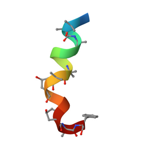
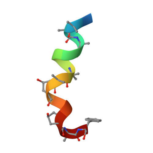
Bergofungin is a peptide antibiotic that is produced by the ascomycetous fungus Emericellopsis donezkii HKI 0059 and belongs to peptaibol subfamily 2. The crystal structure of bergofungin A has been determined and refined to 0.84 Å resolution. This is the second crystal structure of a natural 15-residue peptaibol, after that of samarosporin I. The amino-terminal phenylalanine residue in samarosporin I is exchanged to a valine residue in bergofungin A. According to agar diffusion tests, this results in a nearly inactive antibiotic peptide compared with the moderately active samarosporin I. Crystals were obtained from methanol solutions of purified bergofungin mixed with water. Although there are differences in the intramolecular hydrogen-bonding scheme of samarosporin I, the overall folding is very similar for both peptaibols, namely 3 10 -helical at the termini and α-helical in the middle of the molecules. Bergofungin A and samarosporin I molecules are arranged in a similar way in both lattices. However, the packing of bergofungin A exhibits a second solvent channel along the twofold axis. This latter channel occurs in the vicinity of the N-terminus, where the natural substitution resides.
Organizational Affiliation:
IMBB/FORTH, 70013 Heraklion, Crete, Greece.


