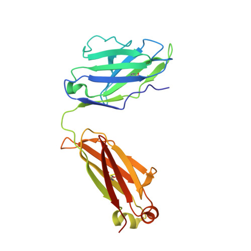
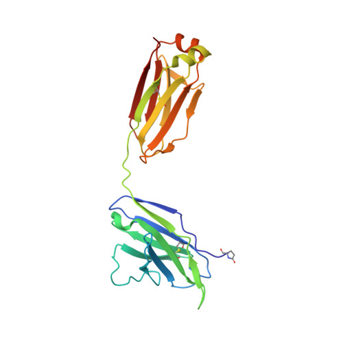
5M6I

Crystal structure of non-cardiotoxic Bence-Jones light chain dimer M8


Experimental Data Snapshot

  • Method: X-RAY DIFFRACTION
  • Resolution: 2.20 Å
  • R-Value Free: 0.227 
  • R-Value Work: 0.195 
  • R-Value Observed: 0.197 

Starting Model: experimental
View more details

wwPDB Validation   3D Report Full Report


This is version 2.2 of the entry. See complete history


Literature

Concurrent structural and biophysical traits link with immunoglobulin light chains amyloid propensity.

Oberti, L.Rognoni, P.Barbiroli, A.Lavatelli, F.Russo, R.Maritan, M.Palladini, G.Bolognesi, M.Merlini, G.Ricagno, S.

(2017) Sci Rep 7: 16809-16809

  • DOI: https://doi.org/10.1038/s41598-017-16953-7
  • Primary Citation of Related Structures:  
    5M6A, 5M6I, 5M76, 5MTL, 5MUD, 5MUH, 5MVG

  • PubMed Abstract: 

    Light chain amyloidosis (AL), the most common systemic amyloidosis, is caused by the overproduction and the aggregation of monoclonal immunoglobulin light chains (LC) in target organs. Due to genetic rearrangement and somatic hypermutation, virtually, each AL patient presents a different amyloidogenic LC. Because of such complexity, the fine molecular determinants of LC aggregation propensity and proteotoxicity are, to date, unclear; significantly, their decoding requires investigating large sets of cases. Aiming to achieve generalizable observations, we systematically characterised a pool of thirteen sequence-diverse full length LCs. Eight amyloidogenic LCs were selected as responsible for severe cardiac symptoms in patients; five non-amyloidogenic LCs were isolated from patients affected by multiple myeloma. Our comprehensive approach (consisting of spectroscopic techniques, limited proteolysis, and X-ray crystallography) shows that low fold stability and high protein dynamics correlate with amyloidogenic LCs, while hydrophobicity, structural rearrangements and nature of the LC dimeric association interface (as observed in seven crystal structures here presented) do not appear to play a significant role in defining amyloid propensity. Based on the structural and biophysical data, our results highlight shared properties driving LC amyloid propensity, and these data will be instrumental for the design of synthetic inhibitors of LC aggregation.


  • Organizational Affiliation

    Dipartimento di Bioscienze, Università degli Studi di Milano, 20133, Milano, Italy.


Macromolecules
Find similar proteins by:  (by identity cutoff)  |  3D Structure
Entity ID: 1
MoleculeChains Sequence LengthOrganismDetailsImage
light chain dimerA [auth B]216Homo sapiensMutation(s): 0 
Entity Groups  
Sequence Clusters30% Identity50% Identity70% Identity90% Identity95% Identity100% Identity
Sequence Annotations
Expand
  • Reference Sequence
Find similar proteins by:  (by identity cutoff)  |  3D Structure
Entity ID: 2
MoleculeChains Sequence LengthOrganismDetailsImage
light chain dimerB [auth A]216Homo sapiensMutation(s): 0 
Entity Groups  
Sequence Clusters30% Identity50% Identity70% Identity90% Identity95% Identity100% Identity
Sequence Annotations
Expand
  • Reference Sequence
Small Molecules
Modified Residues  1 Unique
IDChains TypeFormula2D DiagramParent
PCA
Query on PCA
B [auth A]L-PEPTIDE LINKINGC5 H7 N O3GLN
Experimental Data & Validation

Experimental Data

  • Method: X-RAY DIFFRACTION
  • Resolution: 2.20 Å
  • R-Value Free: 0.227 
  • R-Value Work: 0.195 
  • R-Value Observed: 0.197 
  • Space Group: I 4 2 2
Unit Cell:
Length ( Å )Angle ( ˚ )
a = 229.378α = 90
b = 229.378β = 90
c = 64.429γ = 90
Software Package:
Software NamePurpose
PHENIXrefinement
XDSdata reduction
SCALAdata scaling
PHASERphasing

Structure Validation

View Full Validation Report


Entry History 

Deposition Data

Revision History  (Full details and data files)

  • Version 1.0: 2017-11-15
    Type: Initial release
  • Version 1.1: 2017-12-13
    Changes: Database references
  • Version 2.0: 2020-03-11
    Changes: Atomic model, Polymer sequence
  • Version 2.1: 2024-01-17
    Changes: Advisory, Data collection, Database references, Refinement description
  • Version 2.2: 2024-10-09
    Changes: Structure summary