5M4G | pdb_00005m4g

Crystal Structure of Wild-Type Human Prolidase with Mn ions


Experimental Data Snapshot

  • Method: X-RAY DIFFRACTION
  • Resolution: 1.48 Å
  • R-Value Free: 
    0.167 (Depositor), 0.179 (DCC) 
  • R-Value Work: 
    0.151 (Depositor), 0.164 (DCC) 
  • R-Value Observed: 
    0.151 (Depositor) 

Starting Model: experimental
View more details

wwPDB Validation 3D Report Full Report

Validation slider image for 5M4G

This is version 1.6 of the entry. See complete history

Literature

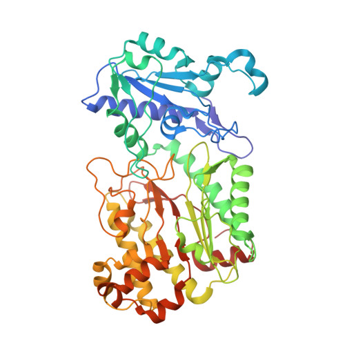
Substrate specificity and reaction mechanism of human prolidase.

Wilk, P.Uehlein, M.Kalms, J.Dobbek, H.Mueller, U.Weiss, M.S.

(2017) FEBS J 284: 2870-2885

  • DOI: https://doi.org/10.1111/febs.14158
  • Primary Citation Related Structures: 
    5M4G, 5M4J, 5M4L, 5M4Q

  • PubMed Abstract: 

    Prolidase is a ubiquitously distributed dipeptidase and the only dipeptidase in humans capable of cleaving the peptide bond preceding the amino acids proline (Pro) or hydroxyproline (Hyp). It is mainly implicated in the degradation of dietary and endogenous proteins. It is also involved in the terminal steps of collagen catabolism by hydrolyzing Pro and Hyp-containing dipeptides. Finally, it is believed to play a role in the regulation of peptidic hormones. Diminished or absent prolidase activity is related to a rare autosomal disease, referred to as prolidase deficiency (PD). This disease manifests itself by a variety of clinical symptoms. To date, there is no definitive cure to PD. This may in part be due to an incomplete understanding of the wild-type (wt) enzyme with respect to substrate-binding mode and consequently the mechanism of the catalyzed reaction. In this work, we describe the high-resolution crystal structures of the wt human prolidase in the ligand-free form as well as in substrate-bound states and in complex with the cleavage product Pro. This series of structures provides much relevant information for the definition of substrate-binding and the reaction mechanism. A recent study on Escherichia coli prolidase revealed how substrates of different length are discriminated. Here, based on our own structural results, we evaluate and extend this analysis. Moreover, we describe and analyze substrate and product binding in the active site and we propose that the crucial catalytic moiety is actually a hydroxide ion. This information significantly advances our understanding of prolidase-based pathologies. The refined structure coordinates as well as the corresponding structure factor amplitudes have been deposited in the PDB under the accession numbers 5M4G, 5M4J, 5M4L, and 5M4Q.


  • Organizational Affiliation
    • Helmholtz-Zentrum Berlin, Macromolecular Crystallography (HZB-MX), Berlin, Germany.

Macromolecule Content 

  • Total Structure Weight: 108.2 kDa 
  • Atom Count: 8,829 
  • Modeled Residue Count: 964 
  • Deposited Residue Count: 966 
  • Unique protein chains: 1

Macromolecules

Find similar proteins by:|  3D Structure
Entity ID: 1
MoleculeChains  Sequence LengthOrganismDetailsImage
Xaa-Pro dipeptidase
A, B
483Homo sapiensMutation(s): 0 
Gene Names: PEPDPRD
EC: 3.4.13.9
UniProt & NIH Common Fund Data Resources
Find proteins for P12955 (Homo sapiens)
Explore P12955 
Go to UniProtKB:  P12955
PHAROS:  P12955
GTEx:  ENSG00000124299 
Entity Groups
Sequence Clusters30% Identity50% Identity70% Identity90% Identity95% Identity100% Identity
UniProt GroupP12955
Sequence Annotations
Expand
Reference Sequence

Small Molecules

Ligands 3 Unique
IDChains Name / Formula / InChI Key2D Diagram3D Interactions
GOL

Query on GOL


Download:Ideal Coordinates CCD File
F [auth A]
G [auth A]
H [auth A]
L [auth B]
M [auth B]
F [auth A],
G [auth A],
H [auth A],
L [auth B],
M [auth B],
N [auth B],
O [auth B]
GLYCEROL
C3 H8 O3
PEDCQBHIVMGVHV-UHFFFAOYSA-N
MN

Query on MN


Download:Ideal Coordinates CCD File
C [auth A],
D [auth A],
I [auth B],
J [auth B]
MANGANESE (II) ION
Mn
WAEMQWOKJMHJLA-UHFFFAOYSA-N
OH

Query on OH


Download:Ideal Coordinates CCD File
E [auth A],
K [auth B]
HYDROXIDE ION
H O
XLYOFNOQVPJJNP-UHFFFAOYSA-M

Experimental Data & Validation

Experimental Data

  • Method: X-RAY DIFFRACTION
  • Resolution: 1.48 Å
  • R-Value Free:  0.167 (Depositor), 0.179 (DCC) 
  • R-Value Work:  0.151 (Depositor), 0.164 (DCC) 
  • R-Value Observed: 0.151 (Depositor) 
Space Group: C 2 2 21
Unit Cell:
Length ( Å )Angle ( ˚ )
a = 103.641α = 90
b = 108.282β = 90
c = 211.452γ = 90
Software Package:
Software NamePurpose
PHENIXrefinement
XSCALEdata scaling
PDB_EXTRACTdata extraction
XDSdata reduction

Structure Validation

View Full Validation Report


Entry History 

Deposition Data

Revision History  (Full details and data files)

  • Version 1.0: 2017-07-12
    Type: Initial release
  • Version 1.1: 2017-07-19
    Changes: Database references
  • Version 1.2: 2017-09-20
    Changes: Database references
  • Version 1.3: 2017-11-29
    Changes: Database references
  • Version 1.4: 2020-04-22
    Changes: Database references
  • Version 1.5: 2024-01-17
    Changes: Data collection, Database references, Derived calculations, Refinement description
  • Version 1.6: 2024-11-06
    Changes: Structure summary