5M3B | pdb_00005m3b

Structure of cobinamide-bound BtuF mutant W66L, the periplasmic vitamin B12 binding protein in E.coli


Experimental Data Snapshot

  • Method: X-RAY DIFFRACTION
  • Resolution: 1.50 Å
  • R-Value Free: 
    0.229 (Depositor), 0.228 (DCC) 
  • R-Value Work: 
    0.196 (Depositor), 0.196 (DCC) 
  • R-Value Observed: 
    0.198 (Depositor) 

Starting Model: experimental
View more details

wwPDB Validation 3D Report Full Report

Validation slider image for 5M3B

Ligand Structure Quality Assessment 


This is version 1.3 of the entry. See complete history

Literature

Conformational Change of a Tryptophan Residue in BtuF Facilitates Binding and Transport of Cobinamide by the Vitamin B12 Transporter BtuCD-F.

Mireku, S.A.Ruetz, M.Zhou, T.Korkhov, V.M.Krautler, B.Locher, K.P.

(2017) Sci Rep 7: 41575-41575

  • DOI: https://doi.org/10.1038/srep41575
  • Primary Citation Related Structures: 
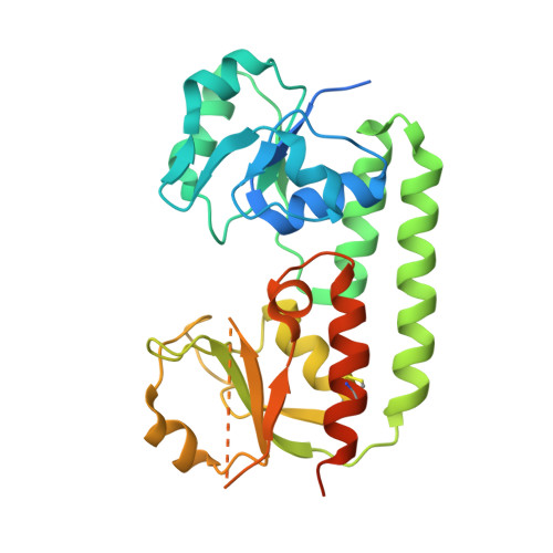
    5M29, 5M2Q, 5M34, 5M3B

  • PubMed Abstract: 

    BtuCD-F is an ABC transporter that mediates cobalamin uptake into Escherichia coli. Early in vivo data suggested that BtuCD-F might also be involved in the uptake of cobinamide, a cobalamin precursor. However, neither was it demonstrated that BtuCD-F indeed transports cobinamide, nor was the structural basis of its recognition known. We synthesized radiolabeled cyano-cobinamide and demonstrated BtuCD-catalyzed in vitro transport, which was ATP- and BtuF-dependent. The crystal structure of cobinamide-bound BtuF revealed a conformational change of a tryptophan residue (W66) in the substrate binding cleft compared to the structure of cobalamin-bound BtuF. High-affinity binding of cobinamide was dependent on W66, because mutation to most other amino acids substantially reduced binding. The structures of three BtuF W66 mutants revealed that tight packing against bound cobinamide was only provided by tryptophan and phenylalanine, in line with the observed binding affinities. In vitro transport rates of cobinamide and cobalamin were not influenced by the substitutions of BtuF W66 under the experimental conditions, indicating that W66 has no critical role in the transport reaction. Our data present the molecular basis of the cobinamide versus cobalamin specificity of BtuCD-F and provide tools for in vitro cobinamide transport and binding assays.


  • Organizational Affiliation
    • Institute of Molecular Biology and Biophysics, Eidgenössische Technische Hochschule (ETH) Zürich, CH-8093 Zürich, Switzerland.

Macromolecule Content 

  • Total Structure Weight: 65.21 kDa 
  • Atom Count: 4,266 
  • Modeled Residue Count: 458 
  • Deposited Residue Count: 578 
  • Unique protein chains: 1

Macromolecules

Find similar proteins by:|  3D Structure
Entity ID: 1
MoleculeChains  Sequence LengthOrganismDetailsImage
Vitamin B12-binding protein
A, B
289Escherichia coli K-12Mutation(s): 1 
Gene Names: btuFyadTb0158JW0154
UniProt
Find proteins for P37028 (Escherichia coli (strain K12))
Explore P37028 
Go to UniProtKB:  P37028
Entity Groups
Sequence Clusters30% Identity50% Identity70% Identity90% Identity95% Identity100% Identity
UniProt GroupP37028
Sequence Annotations
Expand
Reference Sequence

Small Molecules

Ligands 3 Unique
IDChains Name / Formula / InChI Key2D Diagram3D Interactions
CBY

Query on CBY


Download:Ideal Coordinates CCD File
D [auth A],
G [auth B]
COB(II)INAMIDE
C48 H72 Co N11 O8
GFVWZOGCSKVPRA-JFYQDRLCSA-M
GOL

Query on GOL


Download:Ideal Coordinates CCD File
F [auth A],
I [auth B],
J [auth B]
GLYCEROL
C3 H8 O3
PEDCQBHIVMGVHV-UHFFFAOYSA-N
CYN

Query on CYN


Download:Ideal Coordinates CCD File
C [auth A],
E [auth A],
H [auth B]
CYANIDE ION
C N
XFXPMWWXUTWYJX-UHFFFAOYSA-N

Experimental Data & Validation

Experimental Data

  • Method: X-RAY DIFFRACTION
  • Resolution: 1.50 Å
  • R-Value Free:  0.229 (Depositor), 0.228 (DCC) 
  • R-Value Work:  0.196 (Depositor), 0.196 (DCC) 
  • R-Value Observed: 0.198 (Depositor) 
Space Group: C 1 2 1
Unit Cell:
Length ( Å )Angle ( ˚ )
a = 136.28α = 90
b = 90.46β = 110.74
c = 50.81γ = 90
Software Package:
Software NamePurpose
REFMACrefinement
PHENIXrefinement
XSCALEdata scaling
PHENIXphasing

Structure Validation

View Full Validation Report


Ligand Structure Quality Assessment 


Entry History 

Deposition Data

Revision History  (Full details and data files)

  • Version 1.0: 2017-03-01
    Type: Initial release
  • Version 1.1: 2019-10-16
    Changes: Data collection
  • Version 1.2: 2024-01-17
    Changes: Data collection, Database references, Refinement description
  • Version 1.3: 2024-10-23
    Changes: Structure summary