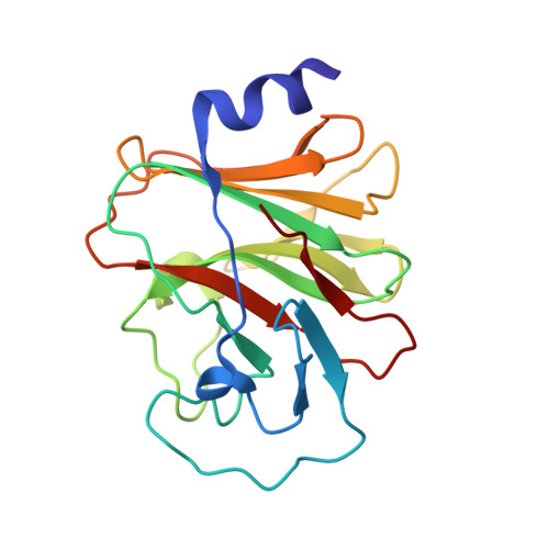
5LYK

CRYSTAL STRUCTURE OF INTRACELLULAR B30.2 DOMAIN OF BTN3A1 BOUND TO CITRATE


Experimental Data Snapshot

  • Method: X-RAY DIFFRACTION
  • Resolution: 1.70 Å
  • R-Value Free: 0.209 
  • R-Value Work: 0.181 
  • R-Value Observed: 0.182 

Starting Model: experimental
View more details

wwPDB Validation   3D Report Full Report


This is version 1.2 of the entry. See complete history


Literature

BTN3A1 Discriminates gamma delta T Cell Phosphoantigens from Nonantigenic Small Molecules via a Conformational Sensor in Its B30.2 Domain.

Salim, M.Knowles, T.J.Baker, A.T.Davey, M.S.Jeeves, M.Sridhar, P.Wilkie, J.Willcox, C.R.Kadri, H.Taher, T.E.Vantourout, P.Hayday, A.Mehellou, Y.Mohammed, F.Willcox, B.E.

(2017) ACS Chem Biol 12: 2631-2643

  • DOI: https://doi.org/10.1021/acschembio.7b00694
  • Primary Citation of Related Structures:  
    5LYG, 5LYK

  • PubMed Abstract: 

    Human Vγ9/Vδ2 T-cells detect tumor cells and microbial infections by recognizing small phosphorylated prenyl metabolites termed phosphoantigens (P-Ag). The type-1 transmembrane protein Butyrophilin 3A1 (BTN3A1) is critical to the P-Ag-mediated activation of Vγ9/Vδ2 T-cells; however, the molecular mechanisms involved in BTN3A1-mediated metabolite sensing are unclear, including how P-Ag's are discriminated from nonantigenic small molecules. Here, we utilized NMR and X-ray crystallography to probe P-Ag sensing by BTN3A1. Whereas the BTN3A1 immunoglobulin variable domain failed to bind P-Ag, the intracellular B30.2 domain bound a range of negatively charged small molecules, including P-Ag, in a positively charged surface pocket. However, NMR chemical shift perturbations indicated BTN3A1 discriminated P-Ag from nonantigenic small molecules by their ability to induce a specific conformational change in the B30.2 domain that propagated from the P-Ag binding site to distal parts of the domain. These results suggest BTN3A1 selectively detects P-Ag intracellularly via a conformational antigenic sensor in its B30.2 domain and have implications for rational design of antigens for Vγ9/Vδ2-based T-cell immunotherapies.


  • Organizational Affiliation

    Institute of Immunology and Immunotherapy, University of Birmingham , Edgbaston, Birmingham, United Kingdom , B15 2TT.


Macromolecules
Find similar proteins by:  (by identity cutoff)  |  3D Structure
Entity ID: 1
MoleculeChains Sequence LengthOrganismDetailsImage
Butyrophilin subfamily 3 member A1187Homo sapiensMutation(s): 0 
Gene Names: BTN3A1BTF5
UniProt & NIH Common Fund Data Resources
Find proteins for O00481 (Homo sapiens)
Explore O00481 
Go to UniProtKB:  O00481
PHAROS:  O00481
GTEx:  ENSG00000026950 
Entity Groups  
Sequence Clusters30% Identity50% Identity70% Identity90% Identity95% Identity100% Identity
UniProt GroupO00481
Sequence Annotations
Expand
  • Reference Sequence
Small Molecules
Ligands 2 Unique
IDChains Name / Formula / InChI Key2D Diagram3D Interactions
FLC
Query on FLC

Download Ideal Coordinates CCD File 
B [auth A]CITRATE ANION
C6 H5 O7
KRKNYBCHXYNGOX-UHFFFAOYSA-K
EDO
Query on EDO

Download Ideal Coordinates CCD File 
C [auth A]1,2-ETHANEDIOL
C2 H6 O2
LYCAIKOWRPUZTN-UHFFFAOYSA-N
Experimental Data & Validation

Experimental Data

  • Method: X-RAY DIFFRACTION
  • Resolution: 1.70 Å
  • R-Value Free: 0.209 
  • R-Value Work: 0.181 
  • R-Value Observed: 0.182 
  • Space Group: P 21 21 2
Unit Cell:
Length ( Å )Angle ( ˚ )
a = 45.5α = 90
b = 125.2β = 90
c = 39.2γ = 90
Software Package:
Software NamePurpose
XSCALEdata scaling
REFMACrefinement
PDB_EXTRACTdata extraction
XDSdata reduction
MOLREPphasing

Structure Validation

View Full Validation Report


Entry History 

Deposition Data

Revision History  (Full details and data files)

  • Version 1.0: 2017-09-13
    Type: Initial release
  • Version 1.1: 2017-11-01
    Changes: Database references
  • Version 1.2: 2024-01-17
    Changes: Advisory, Data collection, Database references, Refinement description