5LWR | pdb_00005lwr

Endothiapepsin in complex with a derivative of fragment 177


Experimental Data Snapshot

  • Method: X-RAY DIFFRACTION
  • Resolution: 1.25 Å
  • R-Value Free: 
    0.131 (Depositor), 0.133 (DCC) 
  • R-Value Work: 
    0.113 (Depositor), 0.114 (DCC) 
  • R-Value Observed: 
    0.114 (Depositor) 

Starting Model: experimental
View more details

wwPDB Validation 3D Report Full Report

Validation slider image for 5LWR

Ligand Structure Quality Assessment 


This is version 1.2 of the entry. See complete history

Literature

A False-Positive Screening Hit in Fragment-Based Lead Discovery: Watch out for the Red Herring.

Cramer, J.Schiebel, J.Wulsdorf, T.Grohe, K.Najbauer, E.E.Ehrmann, F.R.Radeva, N.Zitzer, N.Linne, U.Linser, R.Heine, A.Klebe, G.

(2017) Angew Chem Int Ed Engl 56: 1908-1913

  • DOI: https://doi.org/10.1002/anie.201609824
  • Primary Citation Related Structures: 
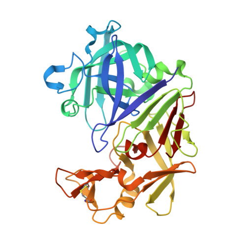
    5LWR, 5LWS, 5LWT, 5LWU

  • PubMed Abstract: 

    With the rising popularity of fragment-based approaches in drug development, more and more attention has to be devoted to the detection of false-positive screening results. In particular, the small size and low affinity of fragments drives screening techniques to their limit. The pursuit of a false-positive hit can cause significant loss of time and resources. Here, we present an instructive and intriguing investigation into the origin of misleading assay results for a fragment that emerged as the most potent binder for the aspartic protease endothiapepsin (EP) across multiple screening assays. This molecule shows its biological effect mainly after conversion into another entity through a reaction cascade that involves major rearrangements of its heterocyclic scaffold. The formed ligand binds EP through an induced-fit mechanism involving remarkable electrostatic interactions. Structural information in the initial screening proved to be crucial for the identification of this false-positive hit.


  • Organizational Affiliation
    • Institut für Pharmazeutische Chemie, Philipps-Universität Marburg, Marbacher Weg 6, 35032, Marburg, Germany.

Macromolecule Content 

  • Total Structure Weight: 35.05 kDa 
  • Atom Count: 2,940 
  • Modeled Residue Count: 330 
  • Deposited Residue Count: 330 
  • Unique protein chains: 1

Macromolecules

Find similar proteins by:|  3D Structure
Entity ID: 1
MoleculeChains  Sequence LengthOrganismDetailsImage
Endothiapepsin330Cryphonectria parasiticaMutation(s): 0 
EC: 3.4.23.22
UniProt
Find proteins for P11838 (Cryphonectria parasitica)
Explore P11838 
Go to UniProtKB:  P11838
Entity Groups
Sequence Clusters30% Identity50% Identity70% Identity90% Identity95% Identity100% Identity
UniProt GroupP11838
Sequence Annotations
Expand
Reference Sequence

Small Molecules

Ligands 5 Unique
IDChains Name / Formula / InChI Key2D Diagram3D Interactions
7B2

Query on 7B2


Download:Ideal Coordinates CCD File
B [auth A]4-[12-[(1-chloranyl-5,6,7-trimethyl-pyrrolo[3,4-d]pyridazin-3-ium-3-yl)methyl]-10,11-dimethyl-3,4,6,7,11-pentazatricyclo[7.3.0.0^{2,6}]dodeca-1(12),2,4,7,9-pentaen-5-yl]-1,2,5-trimethyl-pyrrole-3-carbaldehyde
C27 H29 Cl N9 O
NGGFPHTYSIHBDE-UHFFFAOYSA-N
GOL

Query on GOL


Download:Ideal Coordinates CCD File
C [auth A],
D [auth A],
E [auth A],
K [auth A]
GLYCEROL
C3 H8 O3
PEDCQBHIVMGVHV-UHFFFAOYSA-N
DMS

Query on DMS


Download:Ideal Coordinates CCD File
G [auth A],
H [auth A]
DIMETHYL SULFOXIDE
C2 H6 O S
IAZDPXIOMUYVGZ-UHFFFAOYSA-N
EDO

Query on EDO


Download:Ideal Coordinates CCD File
I [auth A],
J [auth A]
1,2-ETHANEDIOL
C2 H6 O2
LYCAIKOWRPUZTN-UHFFFAOYSA-N
ACT

Query on ACT


Download:Ideal Coordinates CCD File
F [auth A]ACETATE ION
C2 H3 O2
QTBSBXVTEAMEQO-UHFFFAOYSA-M

Experimental Data & Validation

Experimental Data

  • Method: X-RAY DIFFRACTION
  • Resolution: 1.25 Å
  • R-Value Free:  0.131 (Depositor), 0.133 (DCC) 
  • R-Value Work:  0.113 (Depositor), 0.114 (DCC) 
  • R-Value Observed: 0.114 (Depositor) 
Space Group: P 1 21 1
Unit Cell:
Length ( Å )Angle ( ˚ )
a = 45.207α = 90
b = 72.794β = 109.24
c = 52.616γ = 90
Software Package:
Software NamePurpose
PHENIXrefinement
XDSdata reduction
XDSdata scaling
PHASERphasing
Cootmodel building

Structure Validation

View Full Validation Report


Ligand Structure Quality Assessment 


Entry History 

& Funding Information

Deposition Data


Funding OrganizationLocationGrant Number
BMBFGermany05K13RM1

Revision History  (Full details and data files)

  • Version 1.0: 2017-08-09
    Type: Initial release
  • Version 1.1: 2024-01-17
    Changes: Data collection, Database references, Refinement description
  • Version 1.2: 2024-11-20
    Changes: Structure summary