5LUD | pdb_00005lud

Structure of Cyclophilin A in complex with 2,3-Diaminopyridine


Experimental Data Snapshot

  • Method: X-RAY DIFFRACTION
  • Resolution: 1.25 Å
  • R-Value Free: 
    0.220 (Depositor), 0.233 (DCC) 
  • R-Value Work: 
    0.193 (Depositor) 
  • R-Value Observed: 
    0.195 (Depositor) 

Starting Model: experimental
View more details

wwPDB Validation 3D Report Full Report

Validation slider image for 5LUD

This is version 1.2 of the entry. See complete history

Literature

Thermo-kinetic analysis space expansion for cyclophilin-ligand interactions - identification of a new nonpeptide inhibitor using BiacoreTM T200.

Wear, M.A.Nowicki, M.W.Blackburn, E.A.McNae, I.W.Walkinshaw, M.D.

(2017) FEBS Open Bio 7: 533-549

  • DOI: https://doi.org/10.1002/2211-5463.12201
  • Primary Citation Related Structures: 
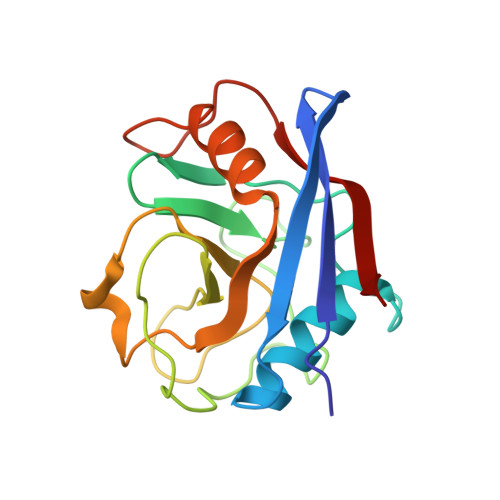
    5LUD

  • PubMed Abstract: 

    We have established a refined methodology for generating surface plasmon resonance sensor surfaces of recombinant his-tagged human cyclophilin-A. Our orientation-specific stabilisation approach captures his-tagged protein under 'physiological conditions' (150 mm NaCl, pH 7.5) and covalently stabilises it on Ni 2+ -nitrilotriacetic acid surfaces, very briefly activated for primary amine-coupling reactions, producing very stable and active surfaces (≥ 95% specific activity) of cyclophilin-A. Variation in protein concentration with the same contact time allows straightforward generation of variable density surfaces, with essentially no loss of activity, making the protocol easily adaptable for studying numerous interactions; from very small fragments, ~ 100 Da, to large protein ligands. This new method results in an increased stability and activity of the immobilised protein and allowed us to expand the thermo-kinetic analysis space, and to determine accurate and robust thermodynamic parameters for the cyclophilin-A-cyclosporin-A interaction. Furthermore, the increased sensitivity of the surface allowed identification of a new nonpeptide inhibitor of cyclophilin-A, from a screen of a fragment library. This fragment, 2,3-diaminopyridine, bound specifically with a mean affinity of 248 ± 60 μm. The X-ray structure of this 109-Da fragment bound in the active site of cyclophilin-A was solved to a resolution of 1.25 Å (PDB: 5LUD), providing new insight into the molecular details for a potential new series of nonpeptide cyclophilin-A inhibitors.


  • Organizational Affiliation
    • The Edinburgh Protein Production Facility (EPPF) Wellcome Trust Centre for Cell Biology (WTCCB) University of Edinburgh UK.

Macromolecule Content 

  • Total Structure Weight: 18.15 kDa 
  • Atom Count: 1,549 
  • Modeled Residue Count: 165 
  • Deposited Residue Count: 165 
  • Unique protein chains: 1

Macromolecules

Find similar proteins by:|  3D Structure
Entity ID: 1
MoleculeChains  Sequence LengthOrganismDetailsImage
Peptidyl-prolyl cis-trans isomerase165Homo sapiensMutation(s): 0 
Gene Names: HEL-S-69p
EC: 5.2.1.8
UniProt & NIH Common Fund Data Resources
Find proteins for P62937 (Homo sapiens)
Explore P62937 
Go to UniProtKB:  P62937
PHAROS:  P62937
GTEx:  ENSG00000196262 
Entity Groups
Sequence Clusters30% Identity50% Identity70% Identity90% Identity95% Identity100% Identity
UniProt GroupP62937
Sequence Annotations
Expand
Reference Sequence

Small Molecules

Ligands 1 Unique
IDChains Name / Formula / InChI Key2D Diagram3D Interactions
76X

Query on 76X


Download:Ideal Coordinates CCD File
B [auth A]pyridine-2,3-diamine
C5 H7 N3
ZZYXNRREDYWPLN-UHFFFAOYSA-N

Experimental Data & Validation

Experimental Data

  • Method: X-RAY DIFFRACTION
  • Resolution: 1.25 Å
  • R-Value Free:  0.220 (Depositor), 0.233 (DCC) 
  • R-Value Work:  0.193 (Depositor) 
  • R-Value Observed: 0.195 (Depositor) 
Space Group: P 21 21 21
Unit Cell:
Length ( Å )Angle ( ˚ )
a = 42.535α = 90
b = 54.477β = 90
c = 86.808γ = 90
Software Package:
Software NamePurpose
REFMACrefinement
XDSdata reduction
Aimlessdata scaling
REFMACphasing

Structure Validation

View Full Validation Report


Entry History 

& Funding Information

Deposition Data


Funding OrganizationLocationGrant Number
Wellcome TrustUnited Kingdom081287/Z/06/Z
Wellcome TrustUnited Kingdom101527/Z/13/Z

Revision History  (Full details and data files)

  • Version 1.0: 2017-04-05
    Type: Initial release
  • Version 1.1: 2017-04-19
    Changes: Database references
  • Version 1.2: 2024-01-17
    Changes: Data collection, Database references, Refinement description