5LU5 | pdb_00005lu5

A quantum half-site enzyme


Experimental Data Snapshot

  • Method: X-RAY DIFFRACTION
  • Resolution: 1.55 Å
  • R-Value Free: 
    0.187 (Depositor), 0.187 (DCC) 
  • R-Value Work: 
    0.161 (Depositor), 0.163 (DCC) 
  • R-Value Observed: 
    0.162 (Depositor) 

wwPDB Validation 3D Report Full Report

Validation slider image for 5LU5

Ligand Structure Quality Assessment 


This is version 1.4 of the entry. See complete history

Literature

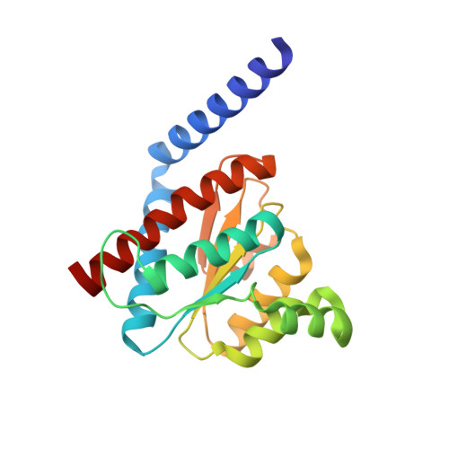
A half-site multimeric enzyme achieves its cooperativity without conformational changes.

Vivoli, M.Pang, J.Harmer, N.J.

(2017) Sci Rep 7: 16529-16529

  • DOI: https://doi.org/10.1038/s41598-017-16421-2
  • Primary Citation Related Structures: 
    5LTZ, 5LU5, 5LU6, 5LU7

  • PubMed Abstract: 

    Cooperativity is a feature many multimeric proteins use to control activity. Here we show that the bacterial heptose isomerase GmhA displays homotropic positive and negative cooperativity among its four protomers. Most similar proteins achieve this through conformational changes: GmhA instead employs a delicate network of hydrogen bonds, and couples pairs of active sites controlled by a unique water channel. This network apparently raises the Lewis acidity of the catalytic zinc, thus increasing the activity at one active site at the cost of preventing substrate from adopting a reactive conformation at the paired negatively cooperative site - a "half-site" behavior. Our study establishes the principle that multimeric enzymes can exploit this cooperativity without conformational changes to maximize their catalytic power and control. More broadly, this subtlety by which enzymes regulate functions could be used to explore new inhibitor design strategies.


  • Organizational Affiliation
    • Department of Biosciences, University of Exeter, Stocker Road, Exeter, EX4 4QD, UK.

Macromolecule Content 

  • Total Structure Weight: 86.3 kDa 
  • Atom Count: 6,799 
  • Modeled Residue Count: 773 
  • Deposited Residue Count: 788 
  • Unique protein chains: 1

Macromolecules

Find similar proteins by:|  3D Structure
Entity ID: 1
MoleculeChains  Sequence LengthOrganismDetailsImage
Phosphoheptose isomerase
A, B, C, D
197Burkholderia pseudomalleiMutation(s): 0 
Gene Names: gmhADP49_467
EC: 5.3.1.28
UniProt
Find proteins for Q93UJ2 (Burkholderia pseudomallei (strain K96243))
Explore Q93UJ2 
Go to UniProtKB:  Q93UJ2
Entity Groups
Sequence Clusters30% Identity50% Identity70% Identity90% Identity95% Identity100% Identity
UniProt GroupQ93UJ2
Sequence Annotations
Expand
Reference Sequence

Small Molecules

Ligands 8 Unique
IDChains Name / Formula / InChI Key2D Diagram3D Interactions
M7P

Query on M7P


Download:Ideal Coordinates CCD File
E [auth A],
I [auth B],
M [auth C],
R [auth D]
7-O-phosphono-D-glycero-alpha-D-manno-heptopyranose
C7 H15 O10 P
SDADNVAZGVDAIM-QTNLNCNHSA-N
PG4

Query on PG4


Download:Ideal Coordinates CCD File
F [auth A],
L [auth B],
S [auth D],
T [auth D]
TETRAETHYLENE GLYCOL
C8 H18 O5
UWHCKJMYHZGTIT-UHFFFAOYSA-N
PGE

Query on PGE


Download:Ideal Coordinates CCD File
H [auth A],
N [auth C],
U [auth D]
TRIETHYLENE GLYCOL
C6 H14 O4
ZIBGPFATKBEMQZ-UHFFFAOYSA-N
PEG

Query on PEG


Download:Ideal Coordinates CCD File
G [auth A]DI(HYDROXYETHYL)ETHER
C4 H10 O3
MTHSVFCYNBDYFN-UHFFFAOYSA-N
GOL

Query on GOL


Download:Ideal Coordinates CCD File
J [auth B],
K [auth B],
O [auth C],
P [auth C]
GLYCEROL
C3 H8 O3
PEDCQBHIVMGVHV-UHFFFAOYSA-N
ACT

Query on ACT


Download:Ideal Coordinates CCD File
W [auth D]ACETATE ION
C2 H3 O2
QTBSBXVTEAMEQO-UHFFFAOYSA-M
MG

Query on MG


Download:Ideal Coordinates CCD File
V [auth D]MAGNESIUM ION
Mg
JLVVSXFLKOJNIY-UHFFFAOYSA-N
NA

Query on NA


Download:Ideal Coordinates CCD File
Q [auth C]SODIUM ION
Na
FKNQFGJONOIPTF-UHFFFAOYSA-N

Experimental Data & Validation

Experimental Data

  • Method: X-RAY DIFFRACTION
  • Resolution: 1.55 Å
  • R-Value Free:  0.187 (Depositor), 0.187 (DCC) 
  • R-Value Work:  0.161 (Depositor), 0.163 (DCC) 
  • R-Value Observed: 0.162 (Depositor) 
Space Group: P 21 21 21
Unit Cell:
Length ( Å )Angle ( ˚ )
a = 73.2α = 90
b = 83.81β = 90
c = 126.78γ = 90
Software Package:
Software NamePurpose
PHENIXrefinement
XDSdata reduction
SCALAdata scaling
MOLREPphasing

Structure Validation

View Full Validation Report


Ligand Structure Quality Assessment 


Entry History 

& Funding Information

Deposition Data


Funding OrganizationLocationGrant Number
Biotechnology and Biological Sciences Research CouncilUnited KingdomBB/H019685/1

Revision History  (Full details and data files)

  • Version 1.0: 2017-12-06
    Type: Initial release
  • Version 1.1: 2017-12-13
    Changes: Database references
  • Version 1.2: 2019-10-16
    Changes: Data collection
  • Version 1.3: 2020-07-29
    Type: Remediation
    Reason: Carbohydrate remediation
    Changes: Derived calculations, Structure summary
  • Version 1.4: 2024-05-08
    Changes: Data collection, Database references, Structure summary