


Crystal structure of nitronate monooxygenase (SO_0471) from Shewanella oneidensis MR-1
Baker, G.E., Race, P.R.To be published.
Experimental Data Snapshot
Starting Model: experimental
View more details
Macromolecule Content
Entity ID: 1 | |||||
|---|---|---|---|---|---|
| Molecule | Chains | Sequence Length | Organism | Details | Image |
| FMN-dependent nitronate monooxygenase | 359 | Shewanella oneidensis MR-1 | Mutation(s): 0 Gene Names: SO_0471 EC: 1.13.12.16 | 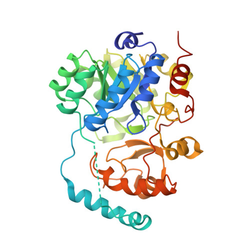 | |
UniProt | |||||
Entity Groups | |||||
| Sequence Clusters | 30% Identity50% Identity70% Identity90% Identity95% Identity100% Identity | ||||
| UniProt Group | Q8EJJ2 | ||||
Sequence AnnotationsExpand | |||||
Reference Sequence | |||||
| Ligands 2 Unique | |||||
|---|---|---|---|---|---|
| ID | Chains | Name / Formula / InChI Key | 2D Diagram | 3D Interactions | |
| FMN Download:Ideal Coordinates CCD File | I [auth A] K [auth B] L [auth C] M [auth D] N [auth E] | FLAVIN MONONUCLEOTIDE C17 H21 N4 O9 P FVTCRASFADXXNN-SCRDCRAPSA-N |  | ||
| GOL Download:Ideal Coordinates CCD File | J [auth A], O [auth E], Q [auth F], S [auth G] | GLYCEROL C3 H8 O3 PEDCQBHIVMGVHV-UHFFFAOYSA-N |  | ||
| Length ( Å ) | Angle ( ˚ ) |
|---|---|
| a = 76.001 | α = 95.31 |
| b = 99.911 | β = 106.23 |
| c = 108.558 | γ = 91.62 |
| Software Name | Purpose |
|---|---|
| REFMAC | refinement |
| Aimless | data scaling |
| PHASER | phasing |
| XDS | data reduction |


