5LR0 | pdb_00005lr0

Binding domain of Botulinum Neurotoxin DC in complex with SialylT


Experimental Data Snapshot

  • Method: X-RAY DIFFRACTION
  • Resolution: 2.59 Å
  • R-Value Free: 
    0.231 (Depositor), 0.233 (DCC) 
  • R-Value Work: 
    0.207 (Depositor), 0.211 (DCC) 
  • R-Value Observed: 
    0.209 (Depositor) 

Starting Model: experimental
View more details

wwPDB Validation 3D Report Full Report

Validation slider image for 5LR0

Ligand Structure Quality Assessment 


This is version 2.1 of the entry. See complete history

Literature

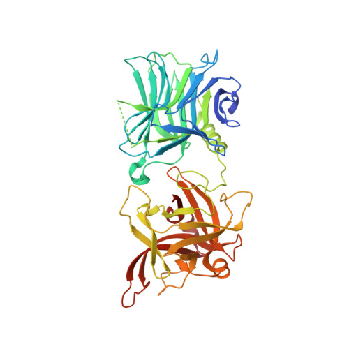
Structural basis for the unique ganglioside and cell membrane recognition mechanism of botulinum neurotoxin DC.

Zhang, S.Berntsson, R.P.Tepp, W.H.Tao, L.Johnson, E.A.Stenmark, P.Dong, M.

(2017) Nat Commun 8: 1637-1637

  • DOI: https://doi.org/10.1038/s41467-017-01534-z
  • Primary Citation Related Structures: 
    5LR0

  • PubMed Abstract: 

    Botulinum neurotoxins (BoNTs), the most potent toxins known, are potential bioterrorism agents. It is well established that all seven serotypes of BoNTs (BoNT/A-G) require complex gangliosides as co-receptors. Here, we report that BoNT/DC, a presumed mosaic toxin between BoNT/D and BoNT/C1, binds and enters efficiently into neurons lacking complex gangliosides and shows no reduction in toxicity in mice deficient in complex gangliosides. The co-crystal structure of BoNT/DC with sialyl-Thomsen-Friedenreich antigen (Sialyl-T) suggests that BoNT/DC recognizes only the sialic acid, but not other moieties in gangliosides. Using liposome flotation assays, we demonstrate that an extended loop in BoNT/DC directly interacts with lipid membranes, and the co-occurring sialic acid binding and loop-membrane interactions mediate the recognition of gangliosides in membranes by BoNT/DC. These findings reveal a unique mechanism for cell membrane recognition and demonstrate that BoNT/DC can use a broad range of sialic acid-containing moieties as co-receptors.


  • Organizational Affiliation
    • Department of Urology, Boston Children's Hospital, Harvard Medical School, Boston, MA, 02115, USA.

Macromolecule Content 

  • Total Structure Weight: 101.5 kDa 
  • Atom Count: 6,944 
  • Modeled Residue Count: 819 
  • Deposited Residue Count: 862 
  • Unique protein chains: 1

Macromolecules

Find similar proteins by:|  3D Structure
Entity ID: 1
MoleculeChains  Sequence LengthOrganismDetailsImage
Botulinum neurotoxin D/C protein
A, B
431Clostridium botulinumMutation(s): 0 
Gene Names: bontboNT DCDCNT
EC: 3.4.24.69
UniProt
Find proteins for A5JGM8 (Clostridium botulinum)
Explore A5JGM8 
Go to UniProtKB:  A5JGM8
Entity Groups
Sequence Clusters30% Identity50% Identity70% Identity90% Identity95% Identity100% Identity
UniProt GroupA5JGM8
Sequence Annotations
Expand
Reference Sequence

Oligosaccharides

Help  
Entity ID: 2
MoleculeChains Length2D Diagram GlycosylationD Interactions
N-acetyl-alpha-neuraminic acid-(2-3)-beta-D-galactopyranose-(1-3)-2-acetamido-2-deoxy-beta-D-galactopyranose
C
3N/A
Glycosylation Resources
GlyTouCan: G02684WR
GlyCosmos: G02684WR
GlyGen: G02684WR
Entity ID: 3
MoleculeChains Length2D Diagram GlycosylationD Interactions
beta-D-galactopyranose-(1-3)-2-acetamido-2-deoxy-beta-D-galactopyranose
D
2N/A
Glycosylation Resources
GlyTouCan: G01534TU
GlyCosmos: G01534TU
GlyGen: G01534TU

Small Molecules

Ligands 1 Unique
IDChains Name / Formula / InChI Key2D Diagram3D Interactions
SIA

Query on SIA


Download:Ideal Coordinates CCD File
E [auth B]N-acetyl-alpha-neuraminic acid
C11 H19 N O9
SQVRNKJHWKZAKO-YRMXFSIDSA-N

Experimental Data & Validation

Experimental Data

  • Method: X-RAY DIFFRACTION
  • Resolution: 2.59 Å
  • R-Value Free:  0.231 (Depositor), 0.233 (DCC) 
  • R-Value Work:  0.207 (Depositor), 0.211 (DCC) 
  • R-Value Observed: 0.209 (Depositor) 
Space Group: P 41 21 2
Unit Cell:
Length ( Å )Angle ( ˚ )
a = 109.699α = 90
b = 109.699β = 90
c = 210.461γ = 90
Software Package:
Software NamePurpose
Aimlessdata scaling
PHASERphasing
REFMACrefinement
PDB_EXTRACTdata extraction
XDSdata reduction

Structure Validation

View Full Validation Report


Ligand Structure Quality Assessment 


Entry History 

Deposition Data

Revision History  (Full details and data files)

  • Version 1.0: 2017-08-30
    Type: Initial release
  • Version 1.1: 2017-12-06
    Changes: Database references
  • Version 2.0: 2020-07-29
    Type: Remediation
    Reason: Carbohydrate remediation
    Changes: Advisory, Atomic model, Data collection, Derived calculations, Structure summary
  • Version 2.1: 2024-01-10
    Changes: Data collection, Database references, Derived calculations, Refinement description, Structure summary