5LP5 | pdb_00005lp5

Complex between Penicillin-Binding Protein (PBP2) and MreC from Helicobacter pylori


Experimental Data Snapshot

  • Method: X-RAY DIFFRACTION
  • Resolution: 2.74 Å
  • R-Value Free: 
    0.292 (Depositor), 0.305 (DCC) 
  • R-Value Work: 
    0.257 (Depositor), 0.267 (DCC) 
  • R-Value Observed: 
    0.258 (Depositor) 

Starting Model: experimental
View more details

wwPDB Validation 3D Report Full Report

Validation slider image for 5LP5

This is version 1.4 of the entry. See complete history

Literature

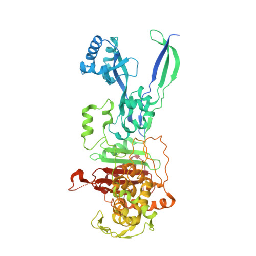
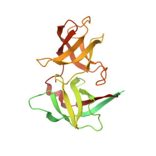
Molecular architecture of the PBP2-MreC core bacterial cell wall synthesis complex.

Contreras-Martel, C.Martins, A.Ecobichon, C.Trindade, D.M.Mattei, P.J.Hicham, S.Hardouin, P.Ghachi, M.E.Boneca, I.G.Dessen, A.

(2017) Nat Commun 8: 776-776

  • DOI: https://doi.org/10.1038/s41467-017-00783-2
  • Primary Citation Related Structures: 
    5LP4, 5LP5

  • PubMed Abstract: 

    Bacterial cell wall biosynthesis is an essential process that requires the coordinated activity of peptidoglycan biosynthesis enzymes within multi-protein complexes involved in cell division (the "divisome") and lateral wall growth (the "elongasome"). MreC is a structural protein that serves as a platform during wall elongation, scaffolding other essential peptidoglycan biosynthesis macromolecules, such as penicillin-binding proteins. Despite the importance of these multi-partite complexes, details of their architecture have remained elusive due to the transitory nature of their interactions. Here, we present the crystal structures of the soluble PBP2:MreC core elongasome complex from Helicobacter pylori, and of uncomplexed PBP2. PBP2 recognizes the two-winged MreC molecule upon opening of its N-terminal region, revealing a hydrophobic zipper that serves as binding platform. The PBP2:MreC interface is essential both for protein recognition in vitro and maintenance of bacterial shape and growth. This work allows visualization as to how peptidoglycan machinery proteins are scaffolded, revealing interaction regions that could be targeted by tailored inhibitors.Bacterial wall biosynthesis is a complex process that requires the coordination of multiple enzymes. Here, the authors structurally characterize the PBP2:MreC complex involved in peptidoglycan elongation and cross-linking, and demonstrate that its disruption leads to loss of H. pylori shape and inability to sustain growth.


  • Organizational Affiliation
    • Univ. Grenoble Alpes, CNRS, CEA, Institut de Biologie Structurale (IBS), Bacterial Pathogenesis Group, F-38000, Grenoble, France.

Macromolecule Content 

  • Total Structure Weight: 247.74 kDa 
  • Atom Count: 13,381 
  • Modeled Residue Count: 1,690 
  • Deposited Residue Count: 2,180 
  • Unique protein chains: 2

Macromolecules

Find similar proteins by:|  3D Structure
Entity ID: 1
MoleculeChains  Sequence LengthOrganismDetailsImage
Penicillin-binding protein 2 (Pbp2)
A, B
594Helicobacter pylori 26695Mutation(s): 0 
Gene Names: HP_1565
UniProt
Find proteins for O26085 (Helicobacter pylori (strain ATCC 700392 / 26695))
Explore O26085 
Go to UniProtKB:  O26085
Entity Groups
Sequence Clusters30% Identity50% Identity70% Identity90% Identity95% Identity100% Identity
UniProt GroupO26085
Sequence Annotations
Expand
Reference Sequence
Find similar proteins by:|  3D Structure
Entity ID: 2
MoleculeChains  Sequence LengthOrganismDetailsImage
Rod shape-determining protein (MreC)
C, D, E, F
248Helicobacter pylori 26695Mutation(s): 0 
Gene Names: HP_1372
UniProt
Find proteins for O25924 (Helicobacter pylori (strain ATCC 700392 / 26695))
Explore O25924 
Go to UniProtKB:  O25924
Entity Groups
Sequence Clusters30% Identity50% Identity70% Identity90% Identity95% Identity100% Identity
UniProt GroupO25924
Sequence Annotations
Expand
Reference Sequence

Experimental Data & Validation

Experimental Data

  • Method: X-RAY DIFFRACTION
  • Resolution: 2.74 Å
  • R-Value Free:  0.292 (Depositor), 0.305 (DCC) 
  • R-Value Work:  0.257 (Depositor), 0.267 (DCC) 
  • R-Value Observed: 0.258 (Depositor) 
Space Group: C 1 2 1
Unit Cell:
Length ( Å )Angle ( ˚ )
a = 338.656α = 90
b = 48.34β = 113.02
c = 151.513γ = 90
Software Package:
Software NamePurpose
REFMACrefinement
XDSdata reduction
XSCALEdata scaling
PHASERphasing

Structure Validation

View Full Validation Report


Entry History 

& Funding Information

Deposition Data


Funding OrganizationLocationGrant Number
French National Research AgencyFrance13-BSV8-0015-01
FAPESPBrazil11/52067-6
European Research CouncilFrance202283

Revision History  (Full details and data files)

  • Version 1.0: 2017-08-23
    Type: Initial release
  • Version 1.1: 2017-08-30
    Changes: Database references, Structure summary
  • Version 1.2: 2017-10-18
    Changes: Database references
  • Version 1.3: 2024-01-10
    Changes: Author supporting evidence, Data collection, Database references, Refinement description
  • Version 1.4: 2024-11-13
    Changes: Structure summary