5LLY | pdb_00005lly

Photosensory Module of Bacteriophytochrome linked Diguanylyl Cyclase from Idiomarina species A28L


Experimental Data Snapshot

  • Method: X-RAY DIFFRACTION
  • Resolution: 2.40 Å
  • R-Value Free: 
    0.230 (Depositor), 0.231 (DCC) 
  • R-Value Work: 
    0.177 (Depositor) 
  • R-Value Observed: 
    0.180 (Depositor) 

Starting Model: experimental
View more details

wwPDB Validation 3D Report Full Report

Validation slider image for 5LLY

Ligand Structure Quality Assessment 


This is version 1.5 of the entry. See complete history

Literature

Long-range allosteric signaling in red light-regulated diguanylyl cyclases.

Gourinchas, G.Etzl, S.Gobl, C.Vide, U.Madl, T.Winkler, A.

(2017) Sci Adv 3: e1602498-e1602498

  • DOI: https://doi.org/10.1126/sciadv.1602498
  • Primary Citation Related Structures: 
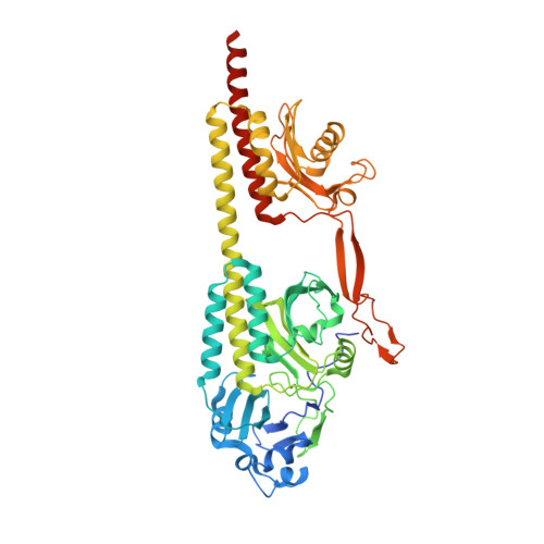
    5LLW, 5LLX, 5LLY

  • PubMed Abstract: 

    Nature has evolved an astonishingly modular architecture of covalently linked protein domains with diverse functionalities to enable complex cellular networks that are critical for cell survival. The coupling of sensory modules with enzymatic effectors allows direct allosteric regulation of cellular signaling molecules in response to diverse stimuli. We present molecular details of red light-sensing bacteriophytochromes linked to cyclic dimeric guanosine monophosphate-producing diguanylyl cyclases. Elucidation of the first crystal structure of a full-length phytochrome with its enzymatic effector, in combination with the characterization of light-induced changes in conformational dynamics, reveals how allosteric light regulation is fine-tuned by the architecture and composition of the coiled-coil sensor-effector linker and also the central helical spine. We anticipate that consideration of molecular principles of sensor-effector coupling, going beyond the length of the characteristic linker, and the appreciation of dynamically driven allostery will open up new directions for the design of novel red light-regulated optogenetic tools.


  • Organizational Affiliation
    • Institute of Biochemistry, Graz University of Technology, Petersgasse 12/II, 8010 Graz, Austria.

Macromolecule Content 

  • Total Structure Weight: 243.35 kDa 
  • Atom Count: 16,833 
  • Modeled Residue Count: 2,013 
  • Deposited Residue Count: 2,116 
  • Unique protein chains: 1

Macromolecules

Find similar proteins by:|  3D Structure
Entity ID: 1
MoleculeChains  Sequence LengthOrganismDetailsImage
Diguanylate cyclase (GGDEF) domain-containing proteinA [auth D],
B [auth A],
C,
D [auth B]
529Idiomarina sp. A28LMutation(s): 0 
Gene Names: A28LD_0430
UniProt
Find proteins for A0ACD6B9P8 (Idiomarina sp. A28L)
Explore A0ACD6B9P8 
Go to UniProtKB:  A0ACD6B9P8
Entity Groups
Sequence Clusters30% Identity50% Identity70% Identity90% Identity95% Identity100% Identity
UniProt GroupA0ACD6B9P8
Sequence Annotations
Expand
Reference Sequence

Small Molecules

Ligands 3 Unique
IDChains Name / Formula / InChI Key2D Diagram3D Interactions
LBV

Query on LBV


Download:Ideal Coordinates CCD File
E [auth D],
H [auth A],
L [auth C],
P [auth B]
3-[2-[(Z)-[3-(2-carboxyethyl)-5-[(Z)-(4-ethenyl-3-methyl-5-oxidanylidene-pyrrol-2-ylidene)methyl]-4-methyl-pyrrol-1-ium -2-ylidene]methyl]-5-[(Z)-[(3E)-3-ethylidene-4-methyl-5-oxidanylidene-pyrrolidin-2-ylidene]methyl]-4-methyl-1H-pyrrol-3- yl]propanoic acid
C33 H37 N4 O6
DKMLMZVDTGOEGU-ISEYCTJISA-O
GOL

Query on GOL


Download:Ideal Coordinates CCD File
K [auth A],
O [auth C]
GLYCEROL
C3 H8 O3
PEDCQBHIVMGVHV-UHFFFAOYSA-N
CL

Query on CL


Download:Ideal Coordinates CCD File
F [auth D]
G [auth D]
I [auth A]
J [auth A]
M [auth C]
F [auth D],
G [auth D],
I [auth A],
J [auth A],
M [auth C],
N [auth C],
Q [auth B],
R [auth B]
CHLORIDE ION
Cl
VEXZGXHMUGYJMC-UHFFFAOYSA-M

Experimental Data & Validation

Experimental Data

  • Method: X-RAY DIFFRACTION
  • Resolution: 2.40 Å
  • R-Value Free:  0.230 (Depositor), 0.231 (DCC) 
  • R-Value Work:  0.177 (Depositor) 
  • R-Value Observed: 0.180 (Depositor) 
Space Group: P 1 21 1
Unit Cell:
Length ( Å )Angle ( ˚ )
a = 83.5α = 90
b = 129β = 96.34
c = 122.01γ = 90
Software Package:
Software NamePurpose
XSCALEdata scaling
PHASERphasing
PHENIXrefinement
PDB_EXTRACTdata extraction
XDSdata reduction

Structure Validation

View Full Validation Report


Ligand Structure Quality Assessment 


Entry History 

& Funding Information

Deposition Data


Funding OrganizationLocationGrant Number
Austrian Science FundAustriaP27124

Revision History  (Full details and data files)

  • Version 1.0: 2017-03-15
    Type: Initial release
  • Version 1.1: 2017-03-22
    Changes: Database references
  • Version 1.2: 2017-08-16
    Changes: Data collection
  • Version 1.3: 2017-09-06
    Changes: Author supporting evidence
  • Version 1.4: 2024-01-10
    Changes: Data collection, Database references, Derived calculations, Refinement description, Structure summary
  • Version 1.5: 2024-10-23
    Changes: Structure summary