5LLS | pdb_00005lls

Porcine dipeptidyl peptidase IV in complex with 8-(3-aminopiperidin-1-yl)-7-[(2-bromophenyl)methyl]-1,3-dimethyl-2,3,6,7-tetrahydro-1H-purine-2,6-dione


Experimental Data Snapshot

  • Method: X-RAY DIFFRACTION
  • Resolution: 2.41 Å
  • R-Value Free: 
    0.228 (Depositor), 0.231 (DCC) 
  • R-Value Work: 
    0.190 (Depositor), 0.195 (DCC) 
  • R-Value Observed: 
    0.192 (Depositor) 

Starting Model: experimental
View more details

wwPDB Validation 3D Report Full Report

Validation slider image for 5LLS

Ligand Structure Quality Assessment 


This is version 2.2 of the entry. See complete history

Literature

Comparative Analysis of Binding Kinetics and Thermodynamics of Dipeptidyl Peptidase-4 Inhibitors and Their Relationship to Structure.

Schnapp, G.Klein, T.Hoevels, Y.Bakker, R.A.Nar, H.

(2016) J Med Chem 59: 7466-7477

  • DOI: https://doi.org/10.1021/acs.jmedchem.6b00475
  • Primary Citation Related Structures: 
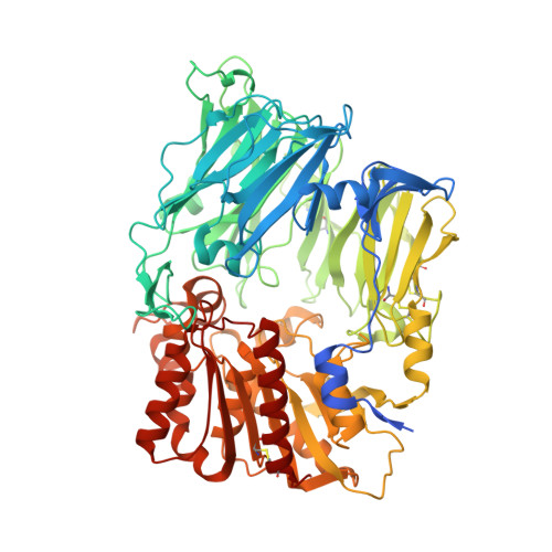
    5LLS

  • PubMed Abstract: 

    The binding kinetics and thermodynamics of dipeptidyl peptidase (DPP)-4 inhibitors (gliptins) were investigated using surface plasmon resonance and isothermal titration calorimetry. Binding of gliptins to DPP-4 is a rapid electrostatically driven process. Off-rates were generally slow partly because of reversible covalent bond formation by some gliptins, and partly because of strong and extensive interactions. Binding of all gliptins is enthalpy-dominated due to strong ionic interactions and strong solvent-shielded hydrogen bonds. Using a congeneric series of molecules which represented the intermediates in the lead optimization program of linagliptin, the onset of slow binding kinetics and development of the thermodynamic repertoire were analyzed in the context of incremental changes of the chemical structures. All compounds rapidly associated, and therefore the optimization of affinity and residence time is highly correlated. The major contributor to the increasing free energy of binding was a strong increase of binding enthalpy, whereas entropic contributions remained low and constant despite significant addition of lipophilicity.


  • Organizational Affiliation
    • Department of Lead Identification and Optimization Support and ‡Department of CardioMetabolic Diseases Research, Boehringer Ingelheim Pharma GmbH & Co. KG , Biberach 88397, Germany.

Macromolecule Content 

  • Total Structure Weight: 364.43 kDa 
  • Atom Count: 25,567 
  • Modeled Residue Count: 2,908 
  • Deposited Residue Count: 3,064 
  • Unique protein chains: 1

Macromolecules

Find similar proteins by:|  3D Structure
Entity ID: 1
MoleculeChains  Sequence LengthOrganismDetailsImage
Dipeptidyl peptidase 4
A, B, C, D
766Sus scrofaMutation(s): 0 
EC: 3.4.14.5
UniProt
Find proteins for P22411 (Sus scrofa)
Explore P22411 
Go to UniProtKB:  P22411
Entity Groups
Sequence Clusters30% Identity50% Identity70% Identity90% Identity95% Identity100% Identity
UniProt GroupP22411
Glycosylation
Glycosylation Sites: 6
Sequence Annotations
Expand
Reference Sequence

Oligosaccharides

Help  
Entity ID: 2
MoleculeChains Length2D Diagram GlycosylationD Interactions
2-acetamido-2-deoxy-beta-D-glucopyranose-(1-4)-2-acetamido-2-deoxy-beta-D-glucopyranose
E, F, G, H, I
E, F, G, H, I, J, K, L, M
2N-Glycosylation
Glycosylation Resources
GlyTouCan: G42666HT
GlyCosmos: G42666HT
GlyGen: G42666HT

Small Molecules

Ligands 3 Unique
IDChains Name / Formula / InChI Key2D Diagram3D Interactions
6Z8

Query on 6Z8


Download:Ideal Coordinates CCD File
AA [auth B]
GA [auth C]
IA [auth C]
LA [auth D]
NA [auth D]
AA [auth B],
GA [auth C],
IA [auth C],
LA [auth D],
NA [auth D],
R [auth A],
T [auth A],
Y [auth B]
8-[(3~{R})-3-azanylpiperidin-1-yl]-7-[(2-bromophenyl)methyl]-1,3-dimethyl-purine-2,6-dione
C19 H23 Br N6 O2
CADOMWLQFMIXFQ-CYBMUJFWSA-N
NAG

Query on NAG


Download:Ideal Coordinates CCD File
BA [auth C]
CA [auth C]
DA [auth C]
EA [auth C]
FA [auth C]
BA [auth C],
CA [auth C],
DA [auth C],
EA [auth C],
FA [auth C],
JA [auth D],
KA [auth D],
N [auth A],
O [auth A],
P [auth A],
Q [auth A],
U [auth B],
V [auth B],
W [auth B],
X [auth B]
2-acetamido-2-deoxy-beta-D-glucopyranose
C8 H15 N O6
OVRNDRQMDRJTHS-FMDGEEDCSA-N
SO4

Query on SO4


Download:Ideal Coordinates CCD File
HA [auth C],
MA [auth D],
S [auth A],
Z [auth B]
SULFATE ION
O4 S
QAOWNCQODCNURD-UHFFFAOYSA-L

Experimental Data & Validation

Experimental Data

  • Method: X-RAY DIFFRACTION
  • Resolution: 2.41 Å
  • R-Value Free:  0.228 (Depositor), 0.231 (DCC) 
  • R-Value Work:  0.190 (Depositor), 0.195 (DCC) 
  • R-Value Observed: 0.192 (Depositor) 
Space Group: P 1
Unit Cell:
Length ( Å )Angle ( ˚ )
a = 61.9α = 112.7
b = 117.68β = 94.6
c = 133.12γ = 91.2
Software Package:
Software NamePurpose
BUSTERrefinement
XDSdata reduction
XSCALEdata scaling
CNXphasing

Structure Validation

View Full Validation Report


Ligand Structure Quality Assessment 


Entry History 

Deposition Data

Revision History  (Full details and data files)

  • Version 1.0: 2016-09-14
    Type: Initial release
  • Version 1.1: 2019-10-16
    Changes: Data collection
  • Version 2.0: 2020-07-29
    Type: Remediation
    Reason: Carbohydrate remediation
    Changes: Atomic model, Data collection, Derived calculations, Structure summary
  • Version 2.1: 2024-01-10
    Changes: Data collection, Database references, Refinement description, Structure summary
  • Version 2.2: 2024-10-23
    Changes: Structure summary