5LLF | pdb_00005llf

Structure of Polyphosphate Kinase 2 mutant D117N from Francisella tularensis with polyphosphate


Experimental Data Snapshot

  • Method: X-RAY DIFFRACTION
  • Resolution: 2.31 Å
  • R-Value Free: 
    0.247 (Depositor), 0.251 (DCC) 
  • R-Value Work: 
    0.198 (Depositor), 0.203 (DCC) 
  • R-Value Observed: 
    0.200 (Depositor) 

Starting Model: experimental
View more details

wwPDB Validation 3D Report Full Report

Validation slider image for 5LLF

Ligand Structure Quality Assessment 


This is version 1.4 of the entry. See complete history

Literature

Substrate recognition and mechanism revealed by ligand-bound polyphosphate kinase 2 structures.

Parnell, A.E.Mordhorst, S.Kemper, F.Giurrandino, M.Prince, J.P.Schwarzer, N.J.Hofer, A.Wohlwend, D.Jessen, H.J.Gerhardt, S.Einsle, O.Oyston, P.C.F.Andexer, J.N.Roach, P.L.

(2018) Proc Natl Acad Sci U S A 115: 3350-3355

  • DOI: https://doi.org/10.1073/pnas.1710741115
  • Primary Citation Related Structures: 
    5LC9, 5LCD, 5LD1, 5LDB, 5LL0, 5LLB, 5LLF, 5MAQ, 5O6K, 5O6M

  • PubMed Abstract: 

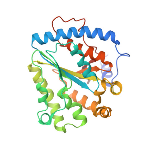
    Inorganic polyphosphate is a ubiquitous, linear biopolymer built of up to thousands of phosphate residues that are linked by energy-rich phosphoanhydride bonds. Polyphosphate kinases of the family 2 (PPK2) use polyphosphate to catalyze the reversible phosphorylation of nucleotide phosphates and are highly relevant as targets for new pharmaceutical compounds and as biocatalysts for cofactor regeneration. PPK2s can be classified based on their preference for nucleoside mono- or diphosphates or both. The detailed mechanism of PPK2s and the molecular basis for their substrate preference is unclear, which is mainly due to the lack of high-resolution structures with substrates or substrate analogs. Here, we report the structural analysis and comparison of a class I PPK2 (ADP-phosphorylating) and a class III PPK2 (AMP- and ADP-phosphorylating), both complexed with polyphosphate and/or nucleotide substrates. Together with complementary biochemical analyses, these define the molecular basis of nucleotide specificity and are consistent with a Mg 2+ catalyzed in-line phosphoryl transfer mechanism. This mechanistic insight will guide the development of PPK2 inhibitors as potential antibacterials or genetically modified PPK2s that phosphorylate alternative substrates.


  • Organizational Affiliation
    • Chemistry, University of Southampton, Southampton, Hampshire SO17 1BJ, United Kingdom.

Macromolecule Content 

  • Total Structure Weight: 132.66 kDa 
  • Atom Count: 9,294 
  • Modeled Residue Count: 1,058 
  • Deposited Residue Count: 1,076 
  • Unique protein chains: 1

Macromolecules

Find similar proteins by:|  3D Structure
Entity ID: 1
MoleculeChains  Sequence LengthOrganismDetailsImage
Polyphosphate kinase 2
A, B, C, D
269Francisella tularensis subsp. tularensis SCHU S4Mutation(s): 1 
Gene Names: ppk2FTT_1564BZ14_1190
EC: 2.7.4.1 (PDB Primary Data), 2.7.4 (UniProt)
UniProt
Find proteins for Q5NEQ5 (Francisella tularensis subsp. tularensis (strain SCHU S4 / Schu 4))
Explore Q5NEQ5 
Go to UniProtKB:  Q5NEQ5
Entity Groups
Sequence Clusters30% Identity50% Identity70% Identity90% Identity95% Identity100% Identity
UniProt GroupQ5NEQ5
Sequence Annotations
Expand
Reference Sequence

Small Molecules

Ligands 5 Unique
IDChains Name / Formula / InChI Key2D Diagram3D Interactions
6YX

Query on 6YX


Download:Ideal Coordinates CCD File
G [auth A][oxidanyl-[oxidanyl-[oxidanyl-[oxidanyl-[oxidanyl-[oxidanyl-[oxidanyl-[oxidanyl-[oxidanyl-[oxidanyl-[oxidanyl-[oxidanyl-[oxidanyl-[oxidanyl-[oxidanyl-[oxidanyl-[oxidanyl-[oxidanyl-[oxidanyl-[oxidanyl-[oxidanyl(phosphonooxy)phosphoryl]oxy-phosphoryl]oxy-phosphoryl]oxy-phosphoryl]oxy-phosphoryl]oxy-phosphoryl]oxy-phosphoryl]oxy-phosphoryl]oxy-phosphoryl]oxy-phosphoryl]oxy-phosphoryl]oxy-phosphoryl]oxy-phosphoryl]oxy-phosphoryl]oxy-phosphoryl]oxy-phosphoryl]oxy-phosphoryl]oxy-phosphoryl]oxy-phosphoryl]oxy-phosphoryl]oxy-phosphoryl] phosphono hydrogen phosphate
H26 O73 P24
YGBPEVYOWOUZHX-UHFFFAOYSA-N
6YY

Query on 6YY


Download:Ideal Coordinates CCD File
J [auth C]bis[oxidanyl-[oxidanyl-[oxidanyl-[oxidanyl(phosphonooxy)phosphoryl]oxy-phosphoryl]oxy-phosphoryl]oxy-phosphoryl] hydrogen phosphate
H13 O34 P11
BWFYHHXRACTZIV-UHFFFAOYSA-N
6YW

Query on 6YW


Download:Ideal Coordinates CCD File
N [auth D][oxidanyl-[oxidanyl-[oxidanyl(phosphonooxy)phosphoryl]oxy-phosphoryl]oxy-phosphoryl] phosphono hydrogen phosphate
H8 O19 P6
LSYVCAOPFHHUHM-UHFFFAOYSA-N
PO4

Query on PO4


Download:Ideal Coordinates CCD File
E [auth A]
F [auth A]
H [auth B]
I [auth B]
L [auth C]
E [auth A],
F [auth A],
H [auth B],
I [auth B],
L [auth C],
M [auth C],
O [auth D],
P [auth D]
PHOSPHATE ION
O4 P
NBIIXXVUZAFLBC-UHFFFAOYSA-K
CL

Query on CL


Download:Ideal Coordinates CCD File
K [auth C]CHLORIDE ION
Cl
VEXZGXHMUGYJMC-UHFFFAOYSA-M

Experimental Data & Validation

Experimental Data

  • Method: X-RAY DIFFRACTION
  • Resolution: 2.31 Å
  • R-Value Free:  0.247 (Depositor), 0.251 (DCC) 
  • R-Value Work:  0.198 (Depositor), 0.203 (DCC) 
  • R-Value Observed: 0.200 (Depositor) 
Space Group: C 2 2 21
Unit Cell:
Length ( Å )Angle ( ˚ )
a = 73.44α = 90
b = 165.94β = 90
c = 255.3γ = 90
Software Package:
Software NamePurpose
PHENIXrefinement
xia2data reduction
xia2data scaling
PHASERphasing

Structure Validation

View Full Validation Report


Ligand Structure Quality Assessment 


Entry History 

& Funding Information

Deposition Data


Funding OrganizationLocationGrant Number
DTRAUnited StatesHDTRA-11-1-007

Revision History  (Full details and data files)

  • Version 1.0: 2017-10-11
    Type: Initial release
  • Version 1.1: 2018-03-21
    Changes: Database references
  • Version 1.2: 2018-04-11
    Changes: Data collection, Database references
  • Version 1.3: 2019-10-16
    Changes: Data collection
  • Version 1.4: 2024-01-10
    Changes: Data collection, Database references, Refinement description