5LKM | pdb_00005lkm

RadA bound to dTDP


Experimental Data Snapshot

  • Method: X-RAY DIFFRACTION
  • Resolution: 3.50 Å
  • R-Value Free: 
    0.224 (Depositor), 0.229 (DCC) 
  • R-Value Work: 
    0.183 (Depositor), 0.184 (DCC) 
  • R-Value Observed: 
    0.185 (Depositor) 

Starting Model: experimental
View more details

wwPDB Validation 3D Report Full Report

Validation slider image for 5LKM

Ligand Structure Quality Assessment 


This is version 1.1 of the entry. See complete history

Literature

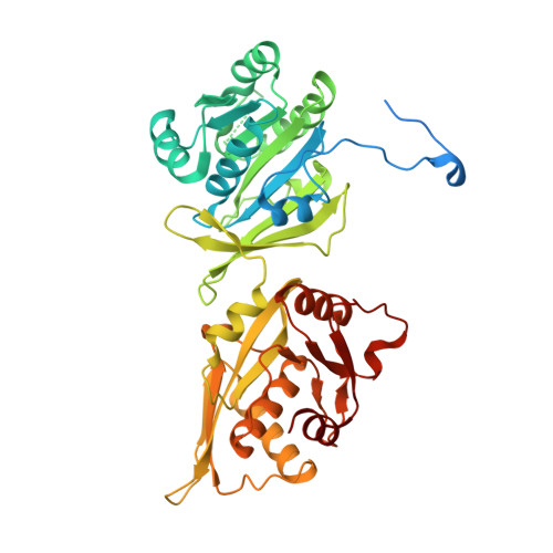
Bacterial RadA is a DnaB-type helicase interacting with RecA to promote bidirectional D-loop extension.

Marie, L.Rapisarda, C.Morales, V.Berge, M.Perry, T.Soulet, A.L.Gruget, C.Remaut, H.Fronzes, R.Polard, P.

(2017) Nat Commun 8: 15638-15638

  • DOI: https://doi.org/10.1038/ncomms15638
  • Primary Citation Related Structures: 
    5LKM, 5LKQ

  • PubMed Abstract: 

    Homologous recombination (HR) is a central process of genome biology driven by a conserved recombinase, which catalyses the pairing of single-stranded DNA (ssDNA) with double-stranded DNA to generate a D-loop intermediate. Bacterial RadA is a conserved HR effector acting with RecA recombinase to promote ssDNA integration. The mechanism of this RadA-mediated assistance to RecA is unknown. Here, we report functional and structural analyses of RadA from the human pathogen Streptococcus pneumoniae. RadA is found to facilitate RecA-driven ssDNA recombination over long genomic distances during natural transformation. RadA is revealed as a hexameric DnaB-type helicase, which interacts with RecA to promote orientated unwinding of branched DNA molecules mimicking D-loop boundaries. These findings support a model of DNA branch migration in HR, relying on RecA-mediated loading of RadA hexamers on each strand of the recipient dsDNA in the D-loop, from which they migrate divergently to facilitate incorporation of invading ssDNA.


  • Organizational Affiliation
    • Laboratoire de Microbiologie et Génétique Moléculaires, UMR5100, Centre de Biologie Intégrative (CBI), Centre National de la Recherche Scientifique (CNRS), Université de Toulouse, UPS, Toulouse F-31062, France.

Macromolecule Content 

  • Total Structure Weight: 149.52 kDa 
  • Atom Count: 8,608 
  • Modeled Residue Count: 1,109 
  • Deposited Residue Count: 1,356 
  • Unique protein chains: 1

Macromolecules

Find similar proteins by:|  3D Structure
Entity ID: 1
MoleculeChains  Sequence LengthOrganismDetailsImage
DNA repair protein RadA
A, B, C
452Streptococcus pneumoniaeMutation(s): 0 
Gene Names: radAERS022181_02072
EC: 3.6.4
UniProt
Find proteins for Q8DRP0 (Streptococcus pneumoniae (strain ATCC BAA-255 / R6))
Explore Q8DRP0 
Go to UniProtKB:  Q8DRP0
Entity Groups
Sequence Clusters30% Identity50% Identity70% Identity90% Identity95% Identity100% Identity
UniProt GroupQ8DRP0
Sequence Annotations
Expand
Reference Sequence

Experimental Data & Validation

Experimental Data

  • Method: X-RAY DIFFRACTION
  • Resolution: 3.50 Å
  • R-Value Free:  0.224 (Depositor), 0.229 (DCC) 
  • R-Value Work:  0.183 (Depositor), 0.184 (DCC) 
  • R-Value Observed: 0.185 (Depositor) 
Space Group: I 2 2 2
Unit Cell:
Length ( Å )Angle ( ˚ )
a = 99.4α = 90
b = 169.9β = 90
c = 187.42γ = 90
Software Package:
Software NamePurpose
REFMACrefinement
BUSTERrefinement
PHENIXrefinement
XDSdata reduction
XDSdata scaling
SHELXDEphasing

Structure Validation

View Full Validation Report


Ligand Structure Quality Assessment 


Entry History 

& Funding Information

Deposition Data


Funding OrganizationLocationGrant Number
European Research CouncilFrance--

Revision History  (Full details and data files)

  • Version 1.0: 2017-06-07
    Type: Initial release
  • Version 1.1: 2024-01-10
    Changes: Author supporting evidence, Data collection, Database references, Derived calculations, Refinement description, Structure summary